 | 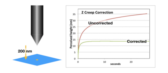 | ||
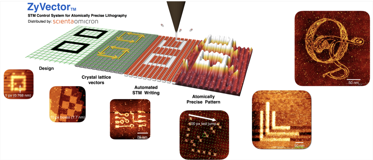
| Zyvex Lab’s ZyVector™ Control system provides the world’s highest (sub-nm) resolution lithography technology. Click here for more information | The Zyvex Creep and Hysteresis Correction Controller. Live tip position control for fast settling times after landing, and precise motion across the surface. Click here for more information. | ||
The 65th International Conference on Electron, Ion and Photon Beam Technology and Nanofabrication
The 27th EIPBN Bizarre/Beautiful Micrograph Contest is now CLOSED. Please see all 2022 award winners and entries below.
The rules include the following:
- Entries have to be of a single image taken with a microscope and should not be significantly altered.
- There is no restriction with respect to the subject matter.
- Electron and ion micrographs have to be black and white.
In 2022, 64 entries were submitted from nine different countries.
The judges were:
- Katherine Cochrane – KLA
- Shelly Muray – Inphora Inc.
- Walter Voit – Adaptive3D
- Sidney Tsai – IBM
There were seven awards:
- Grand Pri(eyes)
- Best Electron Micrograph
- Best Ion Micrograph
- Best Photon Micrograph
- Best Scanning Probe Micrograph
- Most Bizarre
- 3Beamers Choice
There were also 10 Honorable Mention awards given.
To download a PDF with all 64 entries, click HERE
Grand Prize Micrograph
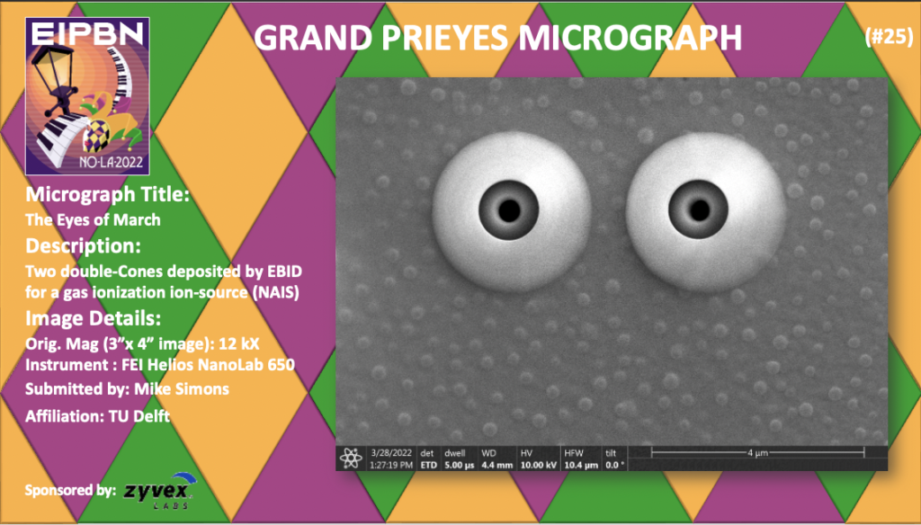
Title: The Eyes of March
Description: Two double-Cones deposited by EBID for a gas ionization ion-source (NAIS)
Magnification (3″ x 4″ image): 12KX
Instrument: FEI Helios NanoLab 650
Submitted by: Mike Simons
Affiliation: TU Delft
Best Electron Micrograph
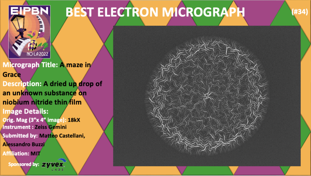
Title: A Maze in Grace
Description: A dried up drop of an unknown substance on niobium nitride thin film
Magnification (3″ x 4″ image): 18KX
Instrument: Zeiss Gemini
Submitted by: Matteo Castellani, Alessandro Buzzi
Affiliation: MIT
Best Ion Micrograph
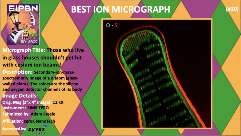
Title: Those Who Live In Glass Houses Shouldn’t Get Hit With Cesium Ion Beams!
Description: Secondary-ion-mass-spectrometry image of a diatom (glass-walled plant). The colors are the silicon and oxygen detector channels of its body
Magnification (3″ x 4″ image): 12KX
Instrument: SIMS:ZERO
Submitted by: Adam Steele
Affiliation: zeroK NanoTech
Best Photon Micrograph
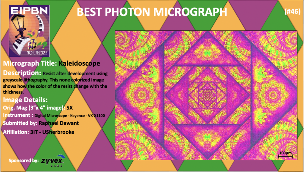
Title: Kaleidoscope
Description: Resist after development using greyscale lithography. This none colorized image shows how the color of the resist change with the thickness.
Magnification (3″ x 4″ image): 5KX
Instrument: : Digital Microscope – Keyence – VK-X1100
Submitted by: Raphael Dawant
Affiliation: 3IT – USherbrooke
Best Scanning Probe Micrograph
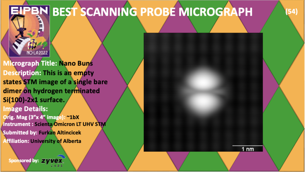
Title: Nano Buns
Description: This is an empty states STM image of a single bare dimer on hydrogen terminated Si(100)-2×1 surface.
Magnification (3″ x 4″ image): ~1bX
Instrument: : Scienta Omicron LT UHV STM
Submitted by: Furkan Altincicek
Affiliation: University of Alberta
Most Bizarre Micrograph

Title: Grumpy McGrumpFace
Description: Material Contrast Image of BiCaCoO sample acquired with AsB detector
Magnification (3″ x 4″ image): 1.82KX
Instrument: Raith, eLINE Plus System (based on Zeiss Gemini SEM)
Submitted by: Frank Nouvertne, Heiner Malchus
Affiliation: GmbH
3-Beamer’s Choice Award
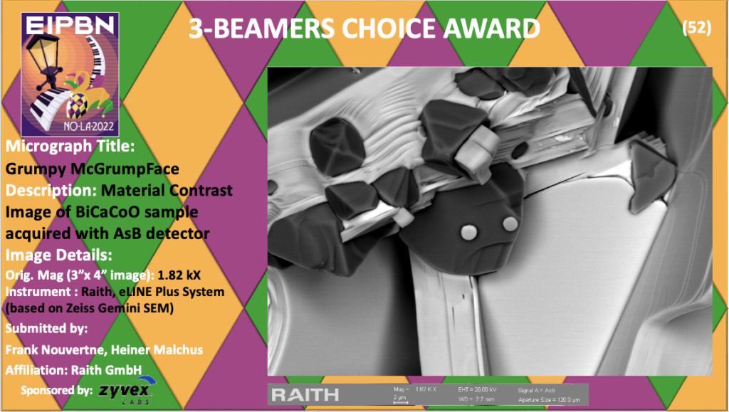
Title: Grumpy McGrumpFace
Description: Material Contrast Image of BiCaCoO sample acquired with AsB detector
Magnification (3″ x 4″ image): 1.82KX
Instrument: Raith, eLINE Plus System (based on Zeiss Gemini SEM)
Submitted by: Frank Nouvertne, Heiner Malchus
Affiliation: GmbH
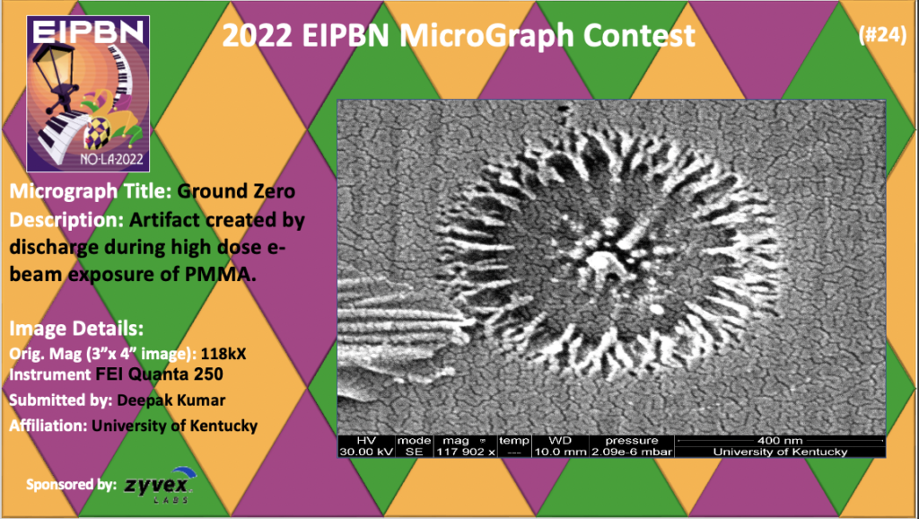
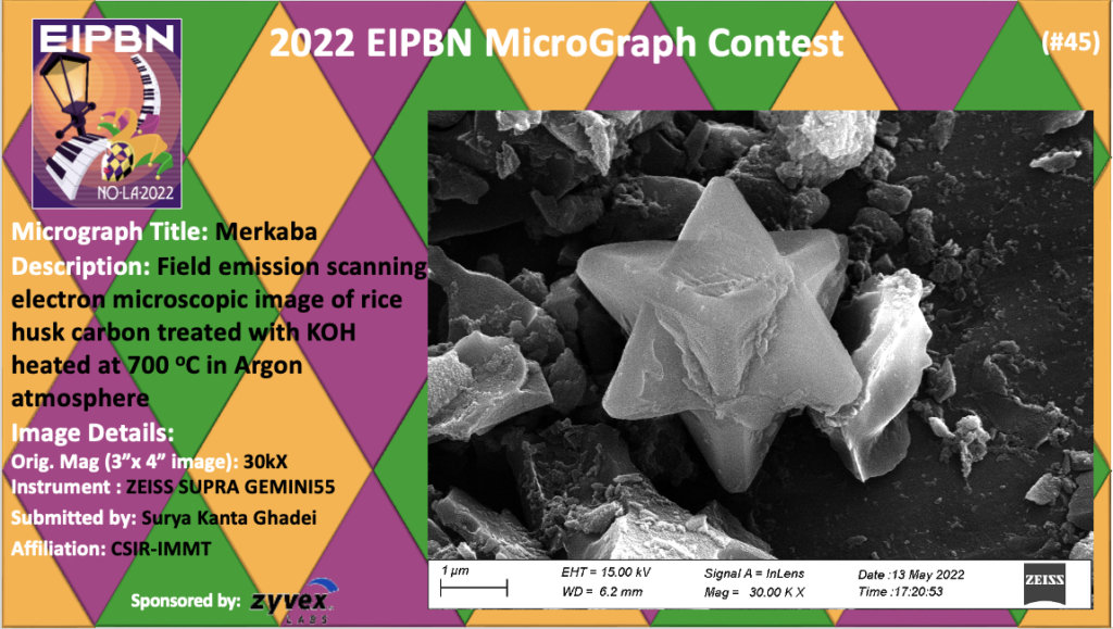
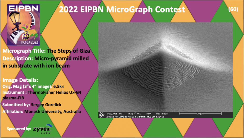
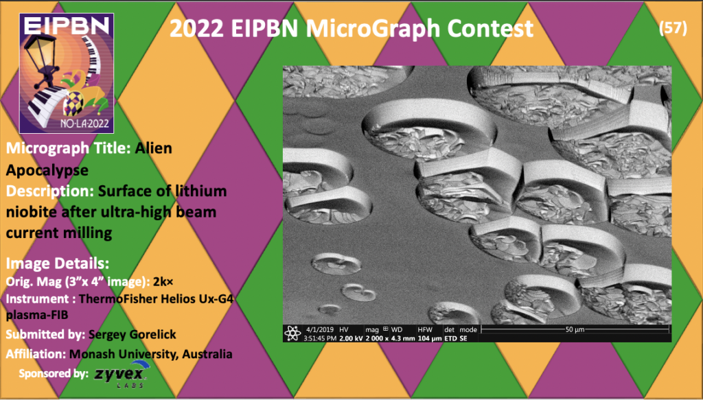
Honorable Mentions