 | 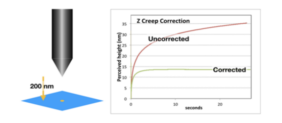 | ||
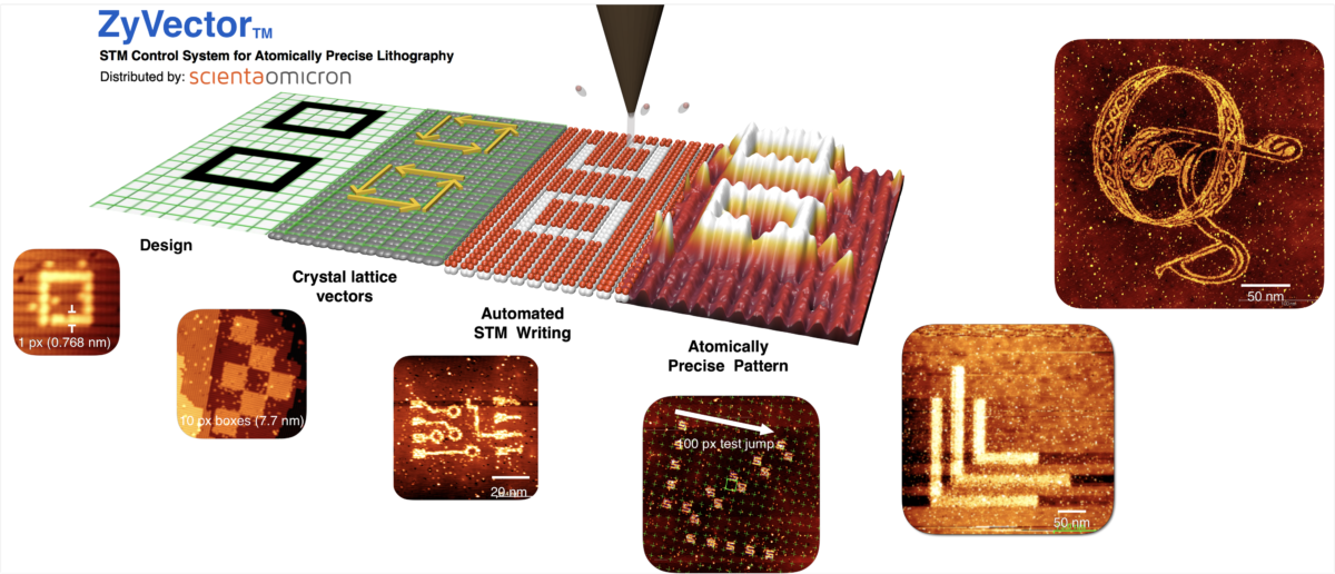
Zyvex Lab’s ZyVector™ Control system provides the world’s highest (sub-nm) resolution lithography technology. Click here for more information | The Zyvex Creep and Hysteresis Correction Controller. Live tip position control for fast settling times after landing, and precise motion across the surface. Click here for more information. |
The 64th International Conference on Electron, Ion and Photon Beam Technology and Nanofabrication
The 26th EIPBN Bizarre/Beautiful Micrograph Contest is now closed. Please see all 2021 award winners and entries below.
The rules include the following:
- Entries have to be of a single image taken with a microscope and should not be significantly altered.
- There is no restriction with respect to the subject matter.
- Electron and ion micrographs have to be black and white.
In 2021, 50 entries were submitted from four different continents
The judges were:
- Chih-Hao Chang – UT Austin
- Qiangfei Xia – U Mass. Amherst
- Alexandra Joshi-Imre – UT Dallas
There were seven awards:
- Grand Prize
- Best Electron Micrograph
- Best Ion Micrograph
- Best Photon Micrograph
- Most Phil-Harmonic Micrograph
- Most Bizarre
- 3Beamers Choice
There were also 12 honorable mention awards given.
To download a PDF with all 50 entries, click HERE
Grand Prize Micrograph
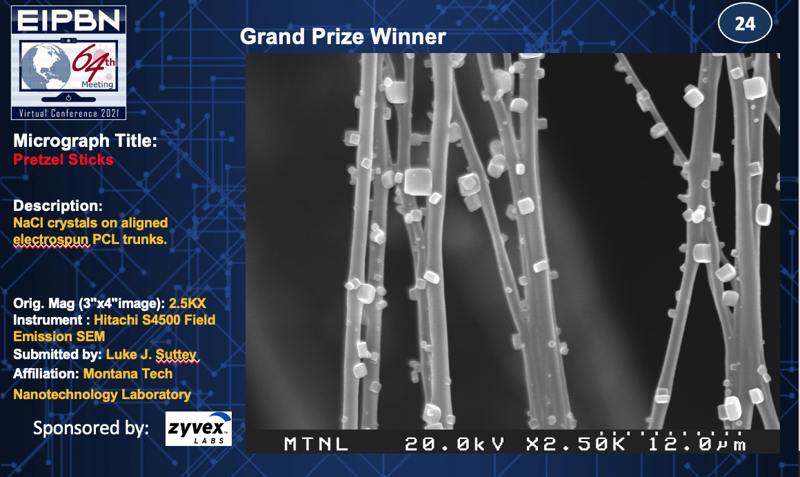
Title: Pretzel Sticks
Description: NaCl crystals on aligned electrospun PCL trunks.
Magnification (3″ x 4″ image): 2.5KX
Instrument: Hitachi S4500 Field Emission SEM
Submitted by: Luke J. Suttey
Affiliation: Montana Tech Nanotechnology Laboratory
Best Electron Micrograph
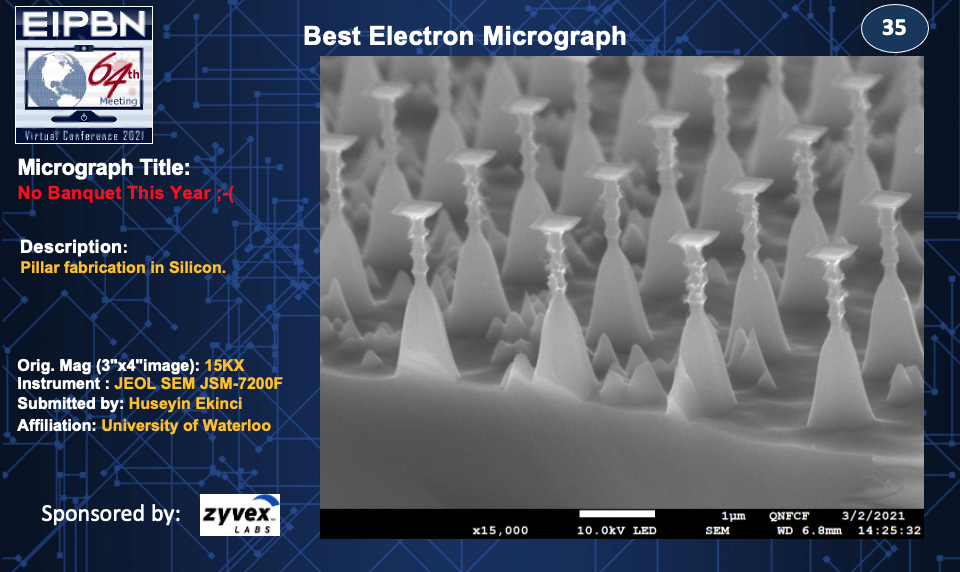
Title: No Banquet This Year ;-(
Description: Pillar fabrication in Silicon.
Magnification (3″ x 4″ image): 15KX
Instrument: JEOL SEM JSM-7200F
Submitted by: Huseyin Ekinci
Affiliation: University of Waterloo
Best Ion Micrograph

Title: No Social Distancing!?
Description: These 1 µm sized structures are fabricated using cryo etch and will be used to puncture the biological cell wall.
Magnification (3″ x 4″ image): 6.6KX
Instrument: FIB FEI Nova 600 NanoLab
Submitted by: Pavani Vamsi & Krishna Nittala
Affiliation: Argonne National Laboratory & The University of Chicago
Best Photon Micrograph
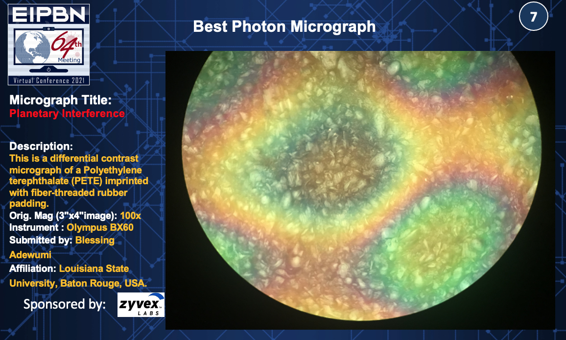
Title: Planetary Interference
Description: This is a differential contrast micrograph of a Polyethylene terephthalate (PETE) imprinted with fiber-threaded rubber padding.
Magnification (3″ x 4″ image): 100x
Instrument: Olympus BX60
Submitted by: Blessing Adewumi
Affiliation: Louisiana State University, Baton Rouge, USA.
Best Scanning Probe Micrograph
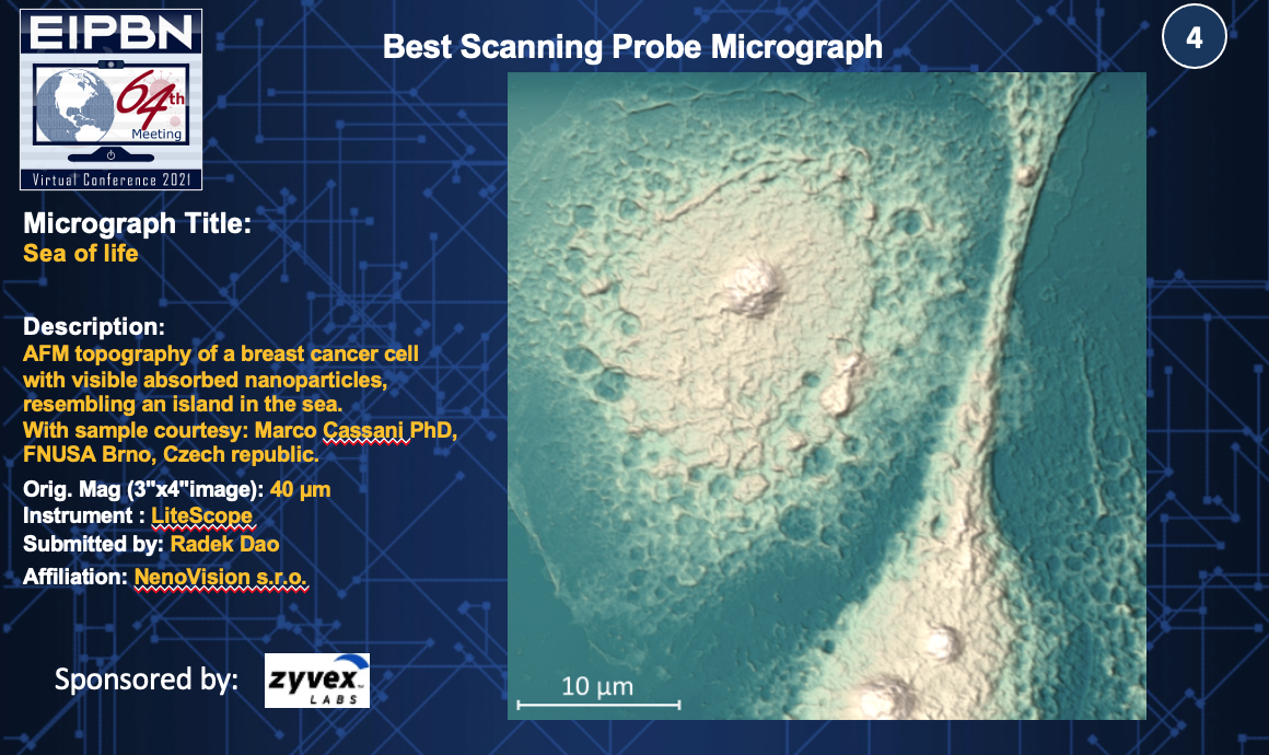
Title: Sea of life
Description: AFM topography of a breast cancer cell with visible absorbed nanoparticles, resembling an island in the sea. Sample courtesy: Marco Cassani PhD, FNUSA Brno, Czech Republic.
Magnification (3″ x 4″ image): 40 µm
Instrument: LiteScope
Submitted by: Radek Dao
Affiliation: NenoVision s.r.o
Most Bizarre Micrograph
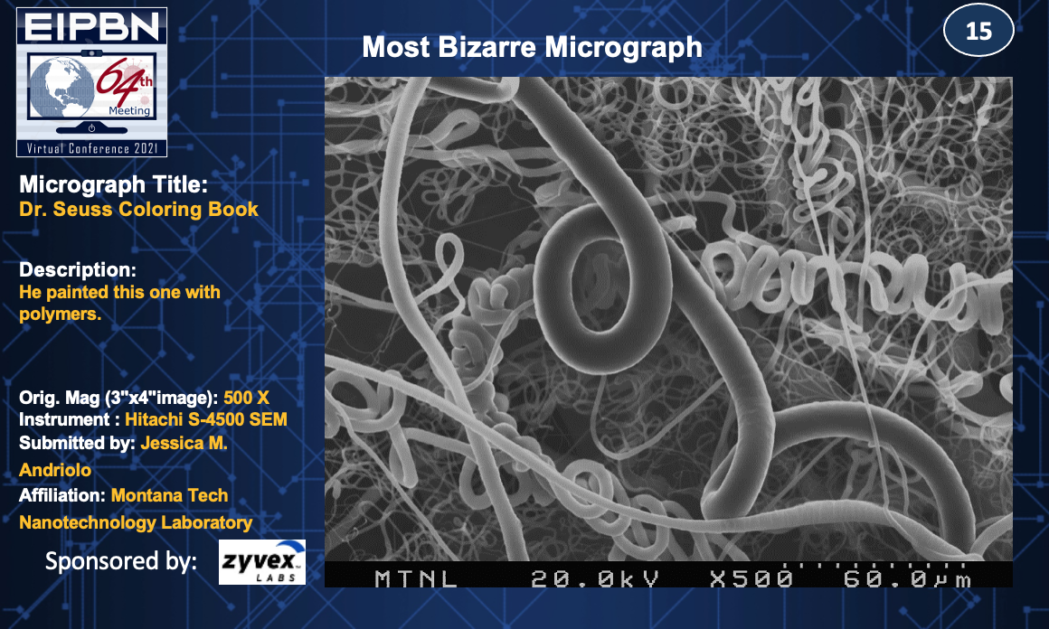
Title: Dr. Seuss Coloring Book
Description: He painted this one with polymers.
Magnification (3″ x 4″ image): 500x
Instrument: Hitachi S-4500 SEM
Submitted by: Jessica M. Andriolo
Affiliation: Montana Tech Nanotechnology Laboratory
3-Beamer’s Choice
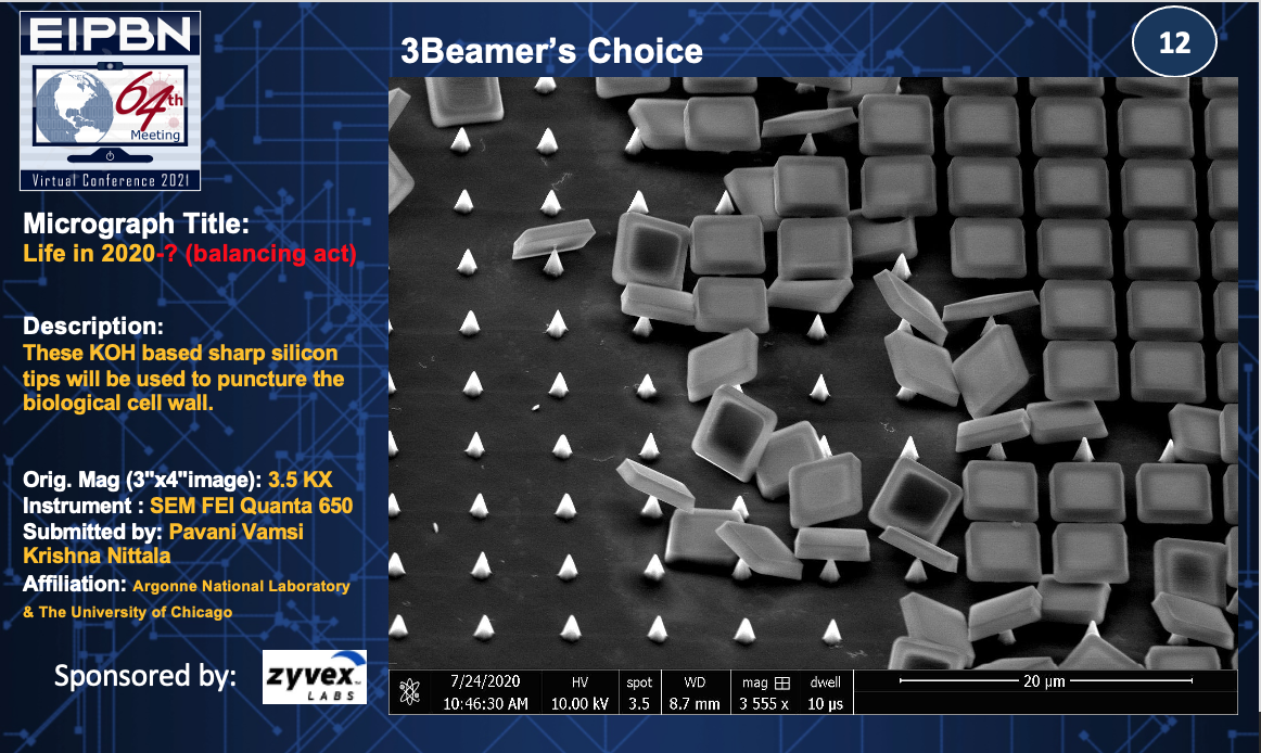
Title: Life in 2020 (Balancing Act)
Description: These KOH based sharp silicon tips will be used to puncture the cell wall.
Magnification (3″x4″ image): 3.5KX
Instrument: SEM FEI Quanta 650
Submitted by: Pavani Vamsi & Krishna Nittala
Affiliation: Argonne National Laboratory & The University of Chicago
Honorable Mentions
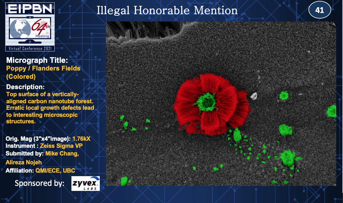
Title: Poppy / Flanders Fields (Colored)
Description: Top surface of a vertically-aligned carbon nanotube forest. Erratic local growth defects lead to interesting microscopic structures.
Magnification (3″x4″ image): 1.76KX
Instrument: Zeiss Sigma VP
Submitted by: Mike Chang & Alireza Nojeh
Affiliation: QMI/ECE, UBC
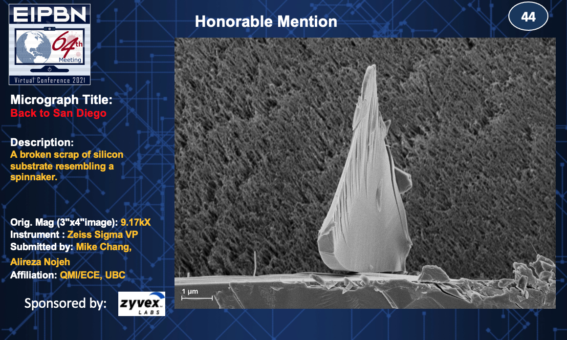
Title: Back to San Diego
Description: A broken scrap of silicon substrate resembling a spinnaker.
Magnification (3″x4″ image): 9.17KX
Instrument: Zeiss Sigma VP
Submitted by: Mike Chang & Alireza Nojeh
Affiliation: QMI/ECE, UBC
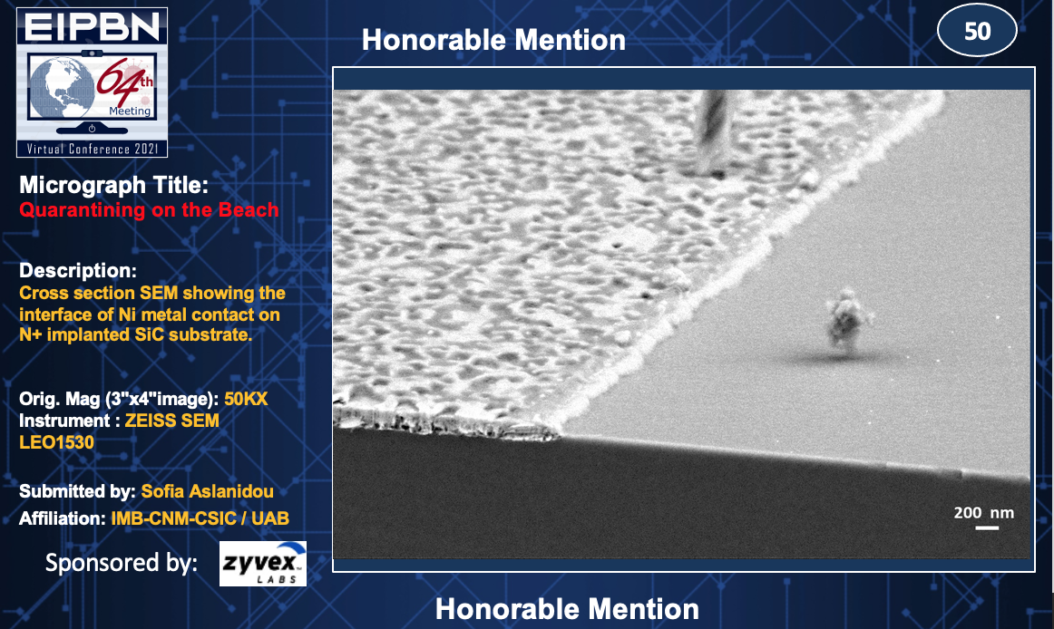
Title: Quarantining on the Beach
Description: Cross section SEM showing the interface of Ni metal contact on N+ implanted SiC substrate.
Magnification (3″x4″ image): 50KX
Instrument: Zeiss SEM LEO 1530
Submitted by: Sofia Aslanidou
Affiliation: IMB-CNM-CSIC / UAB
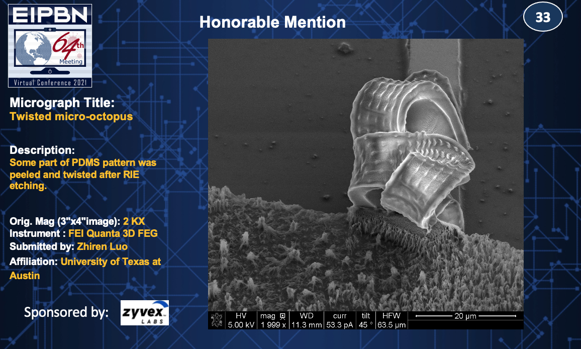
Title: Twisted micro-octopus
Description: Some part of PDMS pattern was peeled and twisted after RIE etching.
Magnification (3″x4″ image): 2KX
Instrument: FEI Quanta 3D FEG
Submitted by: Zhiren Luo
Affiliation: University of Texas at Austin
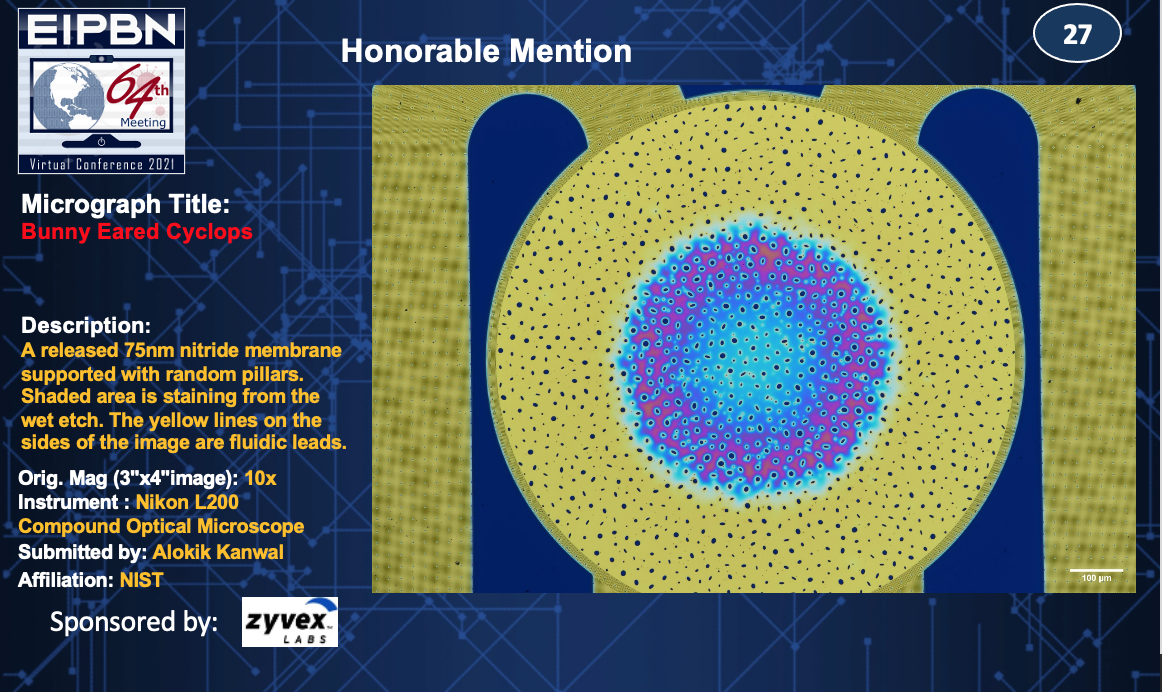
Title: Bunny Eared Cyclops
Description: A released 75nm nitride membrane supported with random pillars. Shaded area is staining from the wet etch. The yellow lines on the sides of the image are fluidic leads.
Magnification (3″x4″ image): 10x
Instrument: Nikon L200 Compound Optical Microscope
Submitted by: Alokik Kanwal
Affiliation: NIST
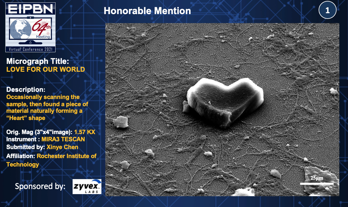
Title: Love For Our World
Description: Occasionally scanning the sample, then found a piece of material naturally forming a “Heart” shape.
Magnification (3″x4″ image): 1.57KX
Instrument: MIRA3 Tescan
Submitted by: Xinye Chen
Affiliation: Rochester Institute of Technology
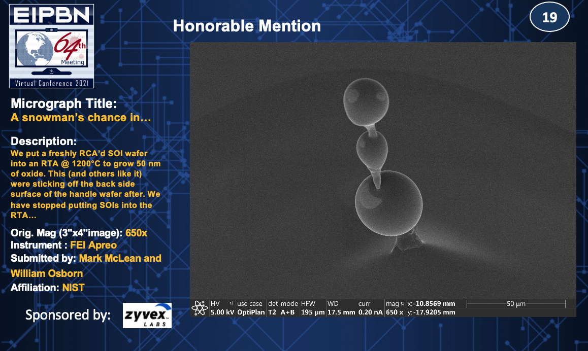
Title: A Snowman’s Chance in…
Description: We put a freshly RCA’d SOI wafer into an RTA @ 1200°C to grow 50nm of oxide. This (and other like it) were sticking off the back side surface of the handle wafer after. We have stopped putting SOIs into the RTA…
Magnification (3″x4″ image): 650x
Instrument: FEI Apreo
Submitted by: Mark McLean and William Osborn
Affiliation: NIST
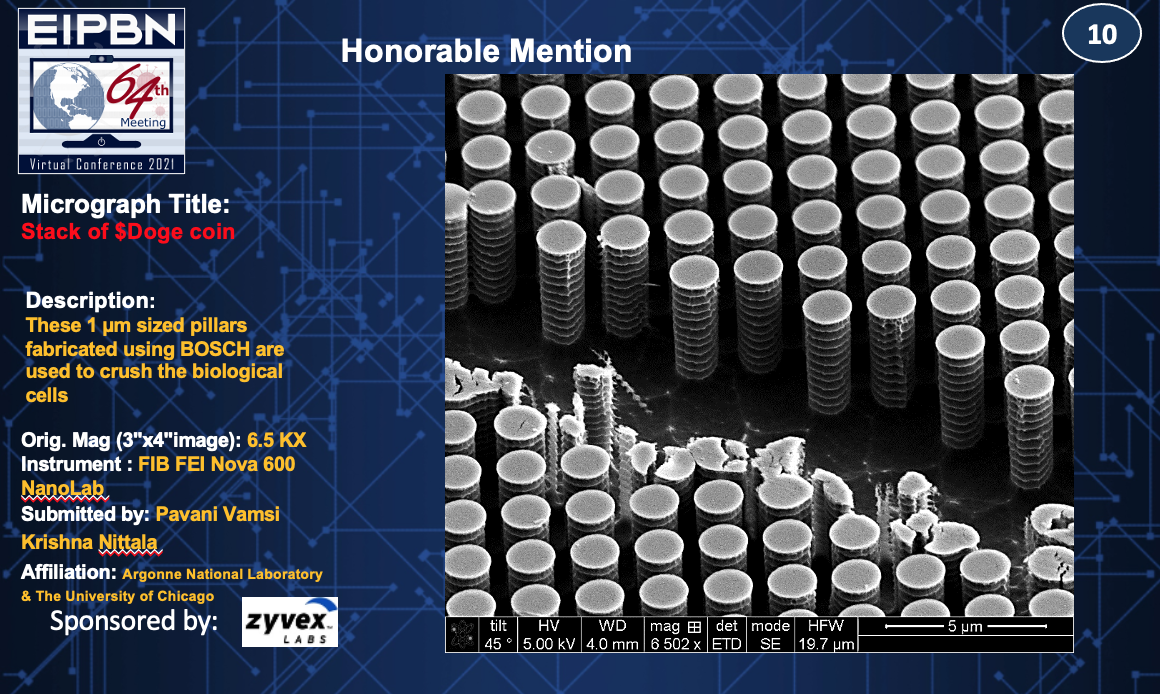
Title: Stack of $Doge Coin
Description: These 1µm sized pillars fabricated using BOSCH are used to crush the biological cells.
Magnification (3″x4″ image): 6.5 KX
Instrument: FIB FEI Nova 600 NanoLab
Submitted by: Pavani Vamsi & Krishna Nittala
Affiliation: Argonne National Laboratory & The University of Chicago
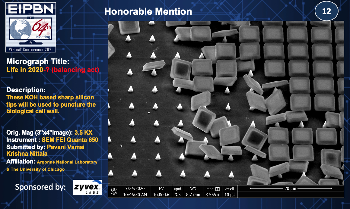
Title: Life in 2020 (A Balancing Act)
Description: These KOH based sharp silicon tips will be used to puncture the biological cell wall.
Magnification (3″x4″ image): 3.5 KX
Instrument: SEM FEI Quanta 650
Submitted by: Pavani Vamsi & Krishna Nittala
Affiliation: Argonne National Laboratory & The University of Chicago
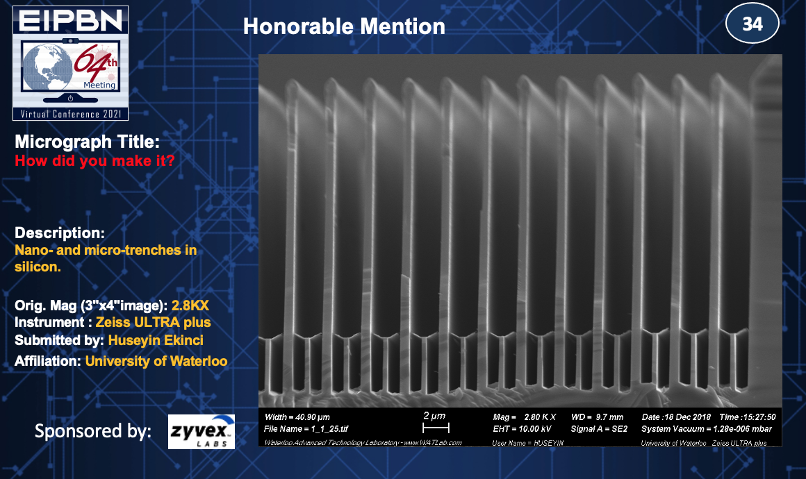
Title: How did you make it?
Description: Nano- and micro-trenches in silicon.
Magnification (3″x4″ image): 2.8KX
Instrument: Zeiss ULTRA plus
Submitted by: Huseyin Ekinci
Affiliation: University of Waterloo
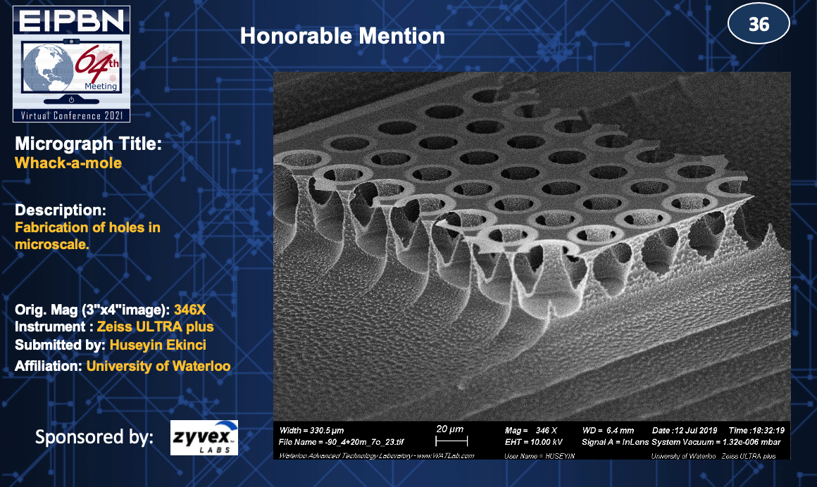
Title: Whack-a-mole
Description: Fabrication of holes in microscale.
Magnification (3″x4″ image): 346X
Instrument: Zeiss ULTRA plus
Submitted by: Huseyin Ekinci
Affiliation: University of Waterloo
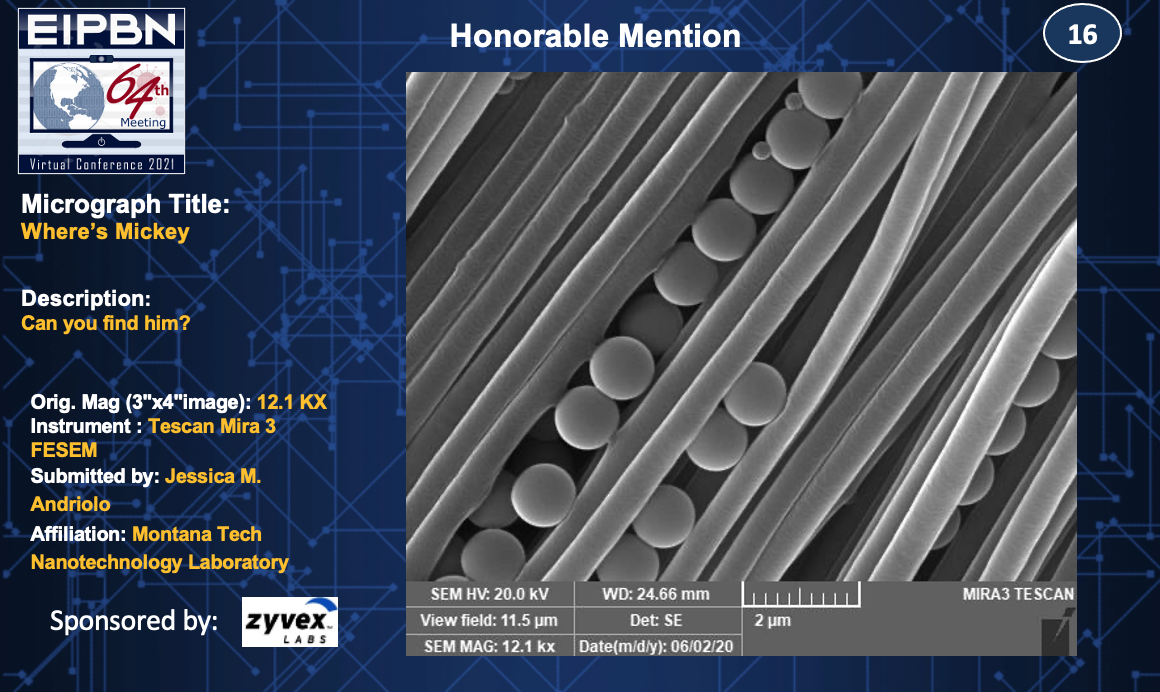
Title: Where’s Mickey
Description: Can you find him?
Magnification (3″x4″ image): 12.1KX
Instrument: Tescan Mira 3 FESEM
Submitted by: Jessica M. Andriolo
Affiliation: Montana Tech Nanotechnology Laboratory