←2009 |
2011→ |
The 54th International Conference on
Electron, Ion and Photon Beam Technology
and Nanofabrication
Bizarre/Beautiful
Micrograph Contest
“ A good Micrograph is worth more than the MegaByte it consumes.”
Entries Presented by Dr. John Randall
The rules include the following:
• Entries have to be of a single image taken with a microscope and could not be significantly altered.
• There is no restriction with respect to the subject matter.
• Electron and ion micrographs have to be black and white.
In 2010, 88 entries were submitted. There were many outstanding micrographs. The work represented in the submitted micrographs covered a wide range of fields including micro mechanical, photonic, and integrated circuit fabrication, chemical and dry etching, carbon nanotube structures, carbon nanotube growth experiments, biological samples, material science experiments and, of course, e-beam, ion beam, and photo lithography experiments.
The panel of judges who selected the award winners were:
• Don Tennant – Cornell
• Edgar Mitchell – Apollo 14 Astronaut
• Cindy Hanson – SPAWAR
There were six awards:
• Grand Prize
• Most Bizzare
• Best Photon Micrograph
• Best Ion Micrograph
• Best Electron Micrograph
• Best Video
There were 17 Honorable Mentions.
All 2010 Entries (with original titles)
Judges exercised their prerogative to liberally interpret the award categories, change the micrograph titles, and even rotate the micrograph if it pleased them.
Title: Alaskan Oasis
Description:Au electroplated structures in defective PMMA mold
Magnification (3″x4″ image): 20 kx
Instrument (Make and Model): SEM Zeiss Supra 55VP
Submitted by: Joan Vila-Comamala
Affiliation: Paul Scherrer Institut (Switzerland)
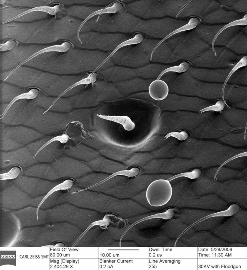
Title: I think it’s down here.
Description:These are the several of the small hairs that are found on the wing of a bee. The scaly nature of the membrane is also seen. Also, several of the hairs show a defect suspected to be a parasite egg.
Magnification (3″x4″ image): 2.4kX
Instrument: Carl Zeiss, ORION Plus (He Ion Microscope)
Submitted by: Shawn McVey and Dave Voci

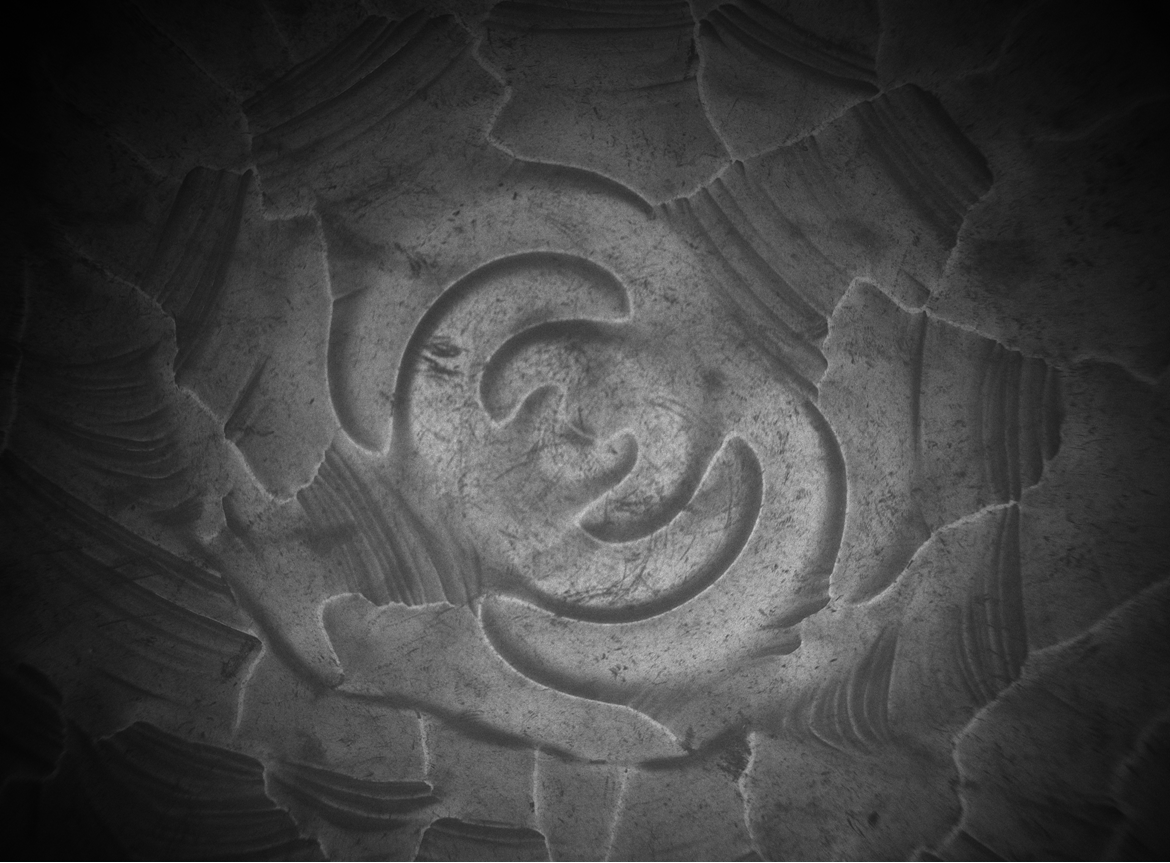
Title: Off Road
Description:This is a bright field optical micrograph of poly(styrene-block-ferrocenyldimethylsilane) (PS-b-PFS) block copolymer thin film. The polymer was spin coated on a thin TEM membrane subjected to hybrid thermal/solvent annealing. This artistic structure appears due to the selective dewetting of the polymer from the thin TEM window (seen in the middle) which oscillates during the spin coating.
Magnification: 100X
Instrument: Zeiss optical microscope
Submitted by: Muruganathan Ramanathan and Seth Darling
Affiliation: Center for Nanoscale Materials, Argonne National Laboratory
Title: Out of Control California Roll
Description:The DIC stack of a hexagonal array of PDMS pillars with magnetic tips. As magnetic tweezer gets close, pillars adhere to each other.
Magnification (3″x4″ image):
Instrument (Make and Model): CCD camera with 40X objective
Submitted by: Saba Ghassemi
Affiliation: Columbia University
Title: Bat Man
Description:Unknown source of contamination on silicon wafer after PMMA resist strip.
Magnification (3″x4″ image): 70x
Instrument (Make and Model): Zeiss Ultra55
Submitted by: Steven Hickman
Affiliation: Cornell University
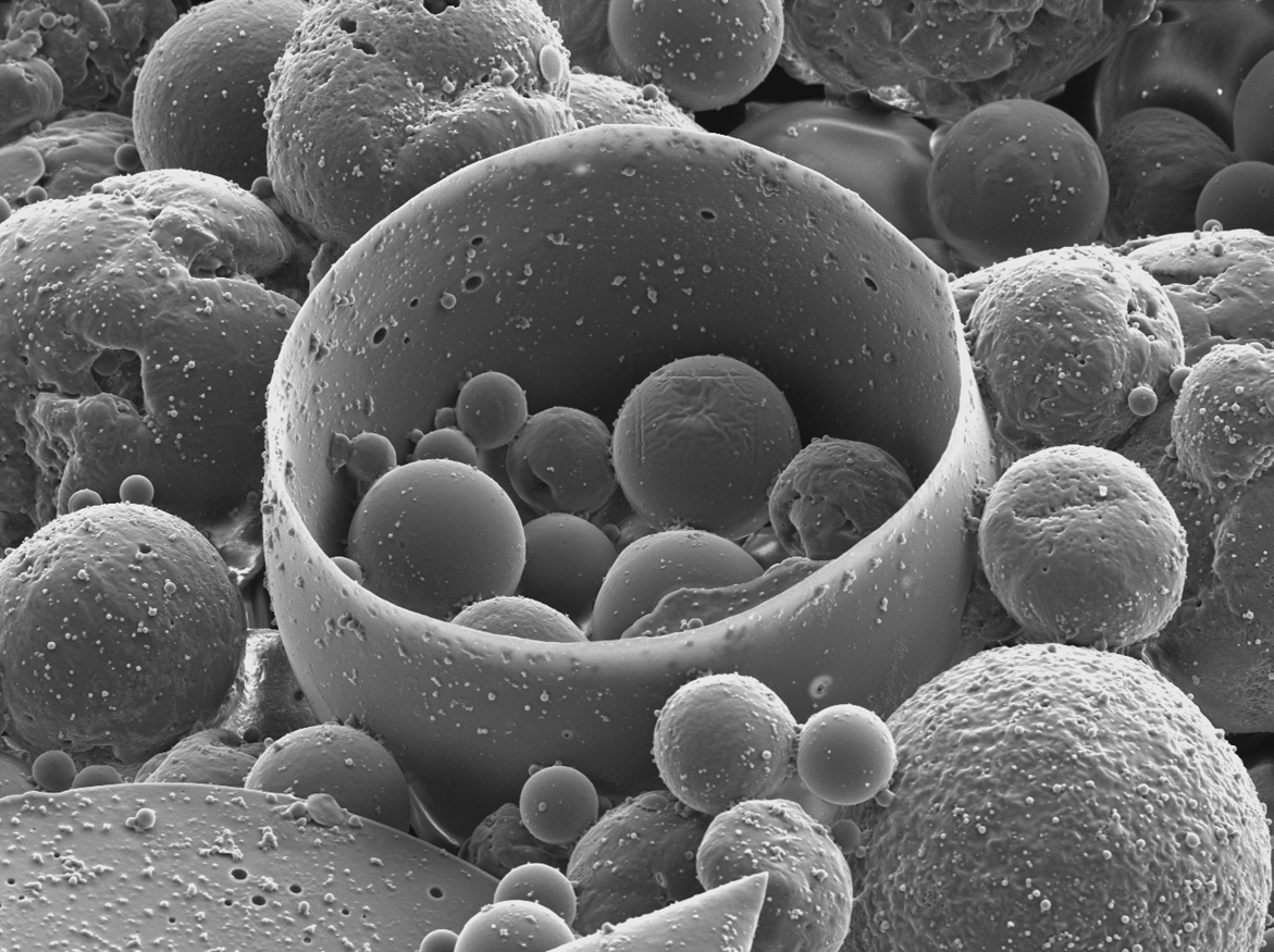
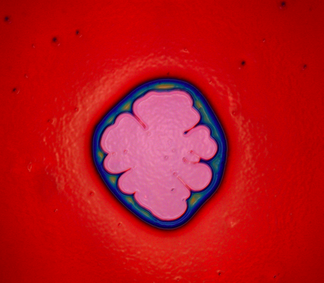
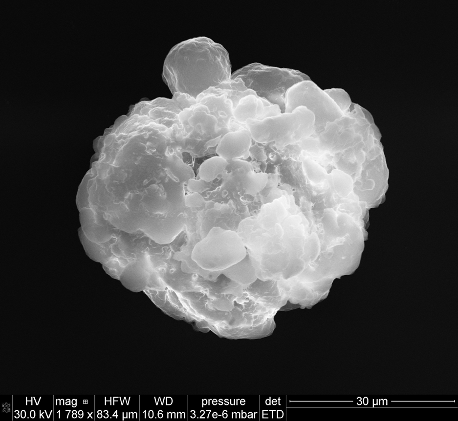
Title: Too Many Eggs for One Basket
Description: Poly(lactic acid) microspheres formed by a W/O/W emulsion and a three leaf clover blade.
Magnification (3″x4″ image): 1000X
instrument (Make and Model): FEI Sirion XL30
Submitted by: Scott Braswell
Affiliation: University of Washington – NTUF
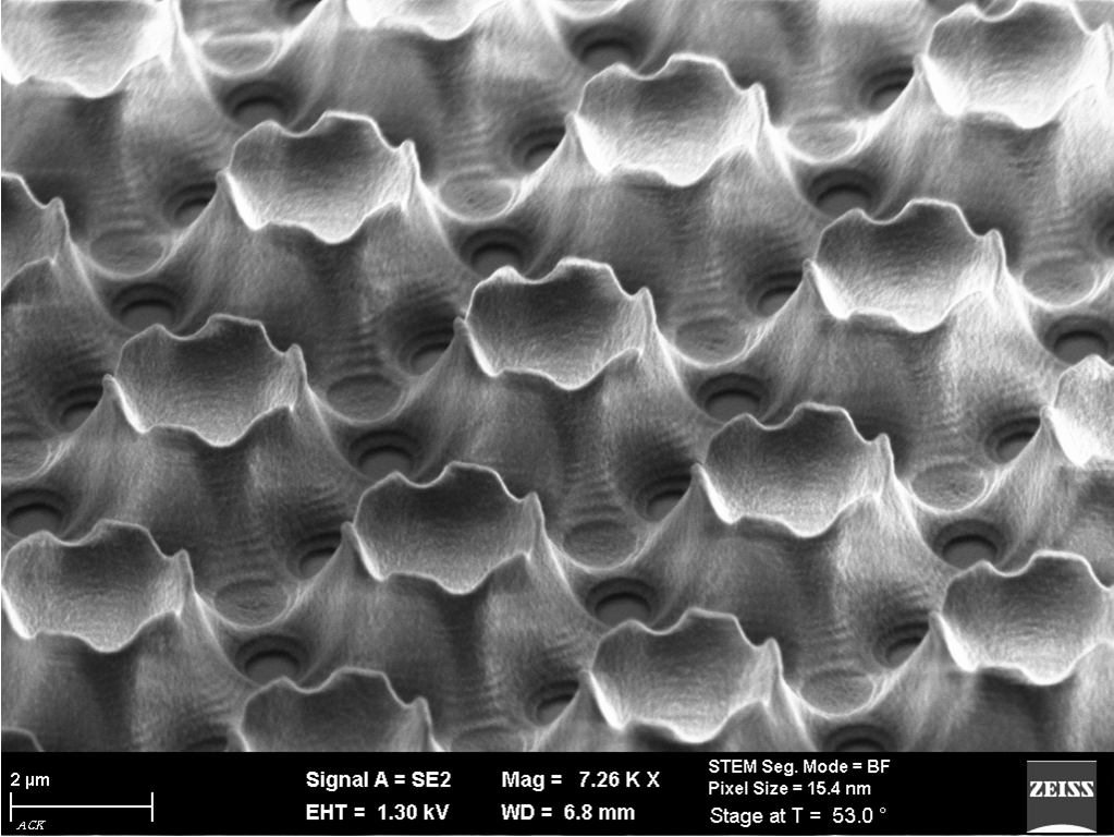
Title: Icelandic Nightmare
Description: Talbot lithography using 1x full field mask aligner with 100 µm exposure gap
Magnification (3″x4″ image): 7260x
Instrument (Make and Model): ZEISS Ultra Plus
Submitted by: Michael Hornung & Uwe Vogler
Affiliation: SUSS MicroTec
HONORABLE MENTION
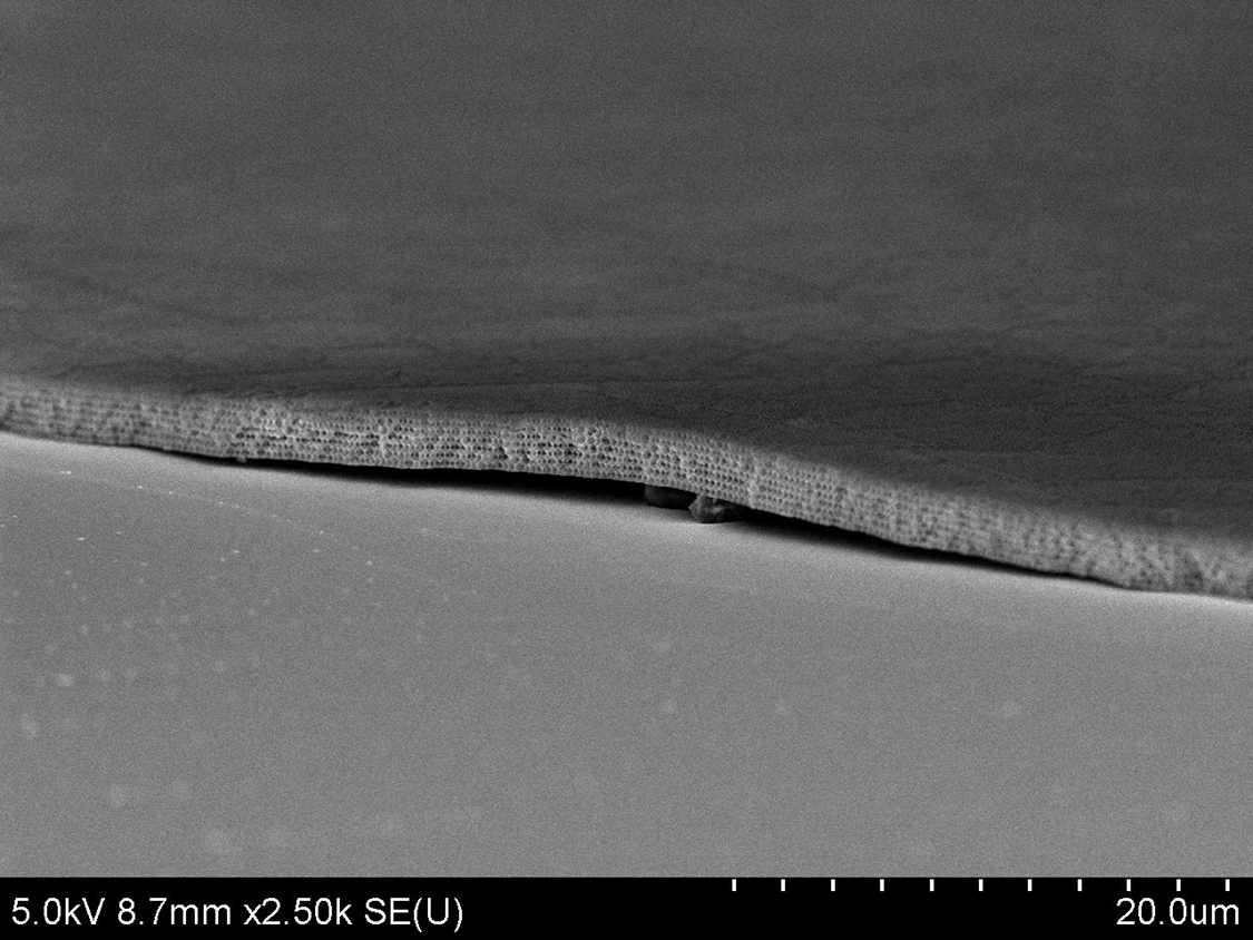
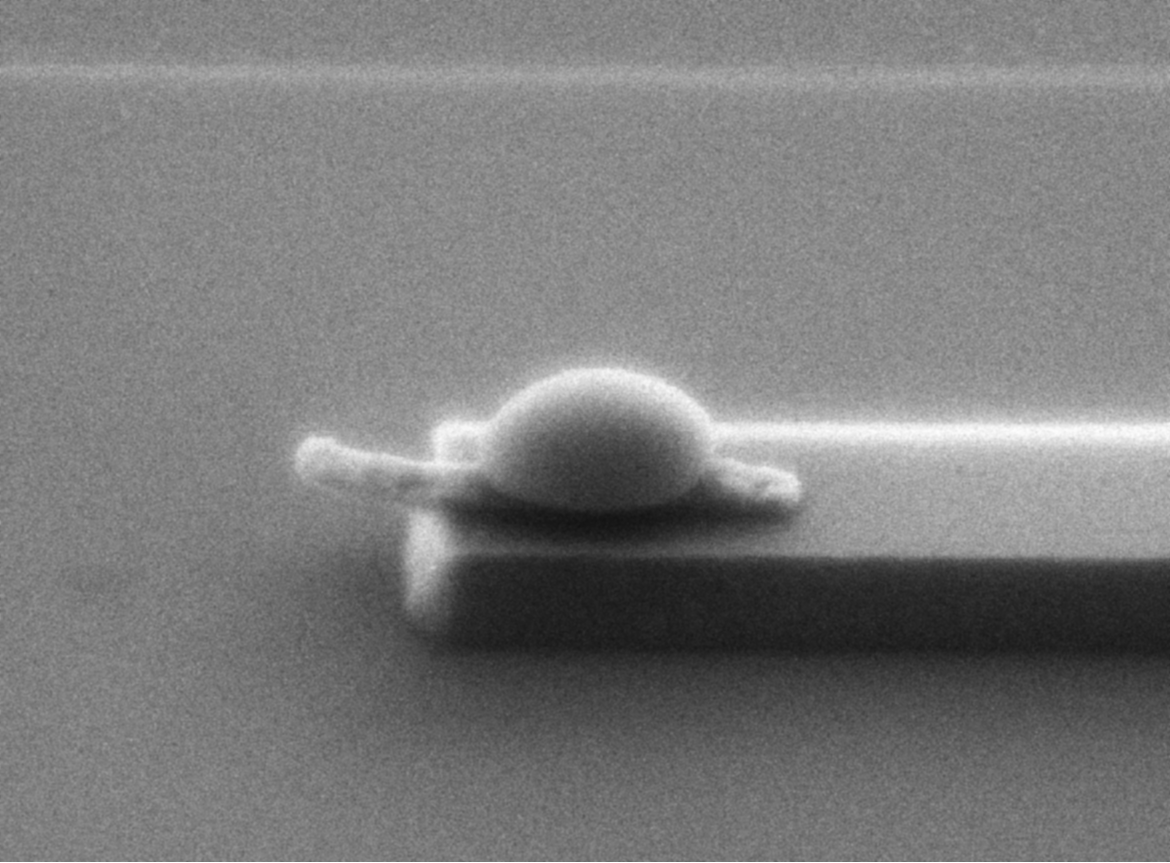
Title: Princess and the Pea
Description: Inverse opal photonic crystal bending over some particles
Magnification (3″x4″ image): 2500X
Instrument (Make and Model): Hitachi S4800 FESEM
Submitted by: Leo Tom Varghese, Li Fan
Affiliation: Birck Nanotechnology Center, Purdue University
HONORABLE MENTION
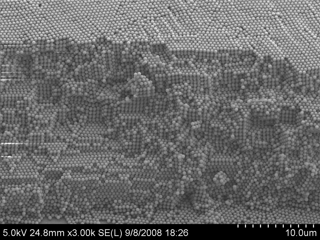
Title: The White Cliffs of Silica
Description: Side view of self assembled silica particles showing 100 crystal orientation
Magnification (3″x4″ image): 3000X
Instrument (Make and Model): Hitachi S4800 FESEM
Submitted by: Leo Tom Varghese, Li Fan
Affiliation: Birck Nanotechnology Center, Purdue University
HONORABLE MENTION
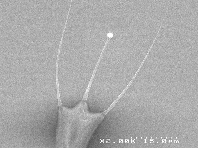
Title: ET Phone Home
Description: SEM picture of a DNA fork fomed on PDMS after evaporation of a DNA solution containing Triton
Magnification (3″x4″ image): X2000
Instrument (Make and Model): SEM Hitachi 4000
Submitted by: J. Cordeiro
Affiliation: BioColloNa LTM CNRS
HONORABLE MENTION
Title: Your Brain on Politics
Description: This is a bright field optical micrograph of poly(styrene-block-ferrocenyldimethylsilane) (PS-b-PFS) block copolymer thin film. Polymer film thickness and the mode of annealing brings out a variety of structures which is currently being explored as an etch mask for mesoscale lithography.
Magnification: 100X
Instrument: Zeiss optical microscope (cross-polarization mode)
Submitted by: Muruganathan Ramanathan and Seth Darling
Affiliation: Center for Nanoscale Materials, Argonne National Laboratory
HONORABLE MENTION
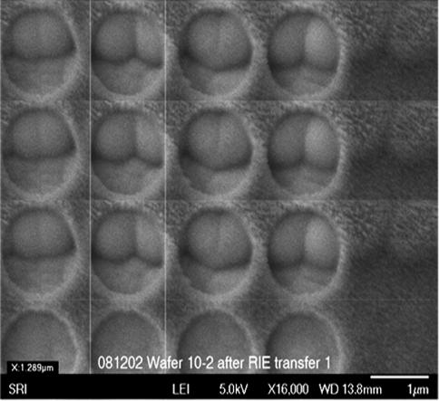
Title: Under the bleachers
Description: Funny bottom structures revealed beneath the upper layer after it was etched out.
Magnification (3″x4″ image): 16kx
Instrument (JEO L JSM-6700)
Submitted by: Yehiel Gotkis & Alan Brodie
Affiliation: KLA-Tencor
HONORABLE MENTION
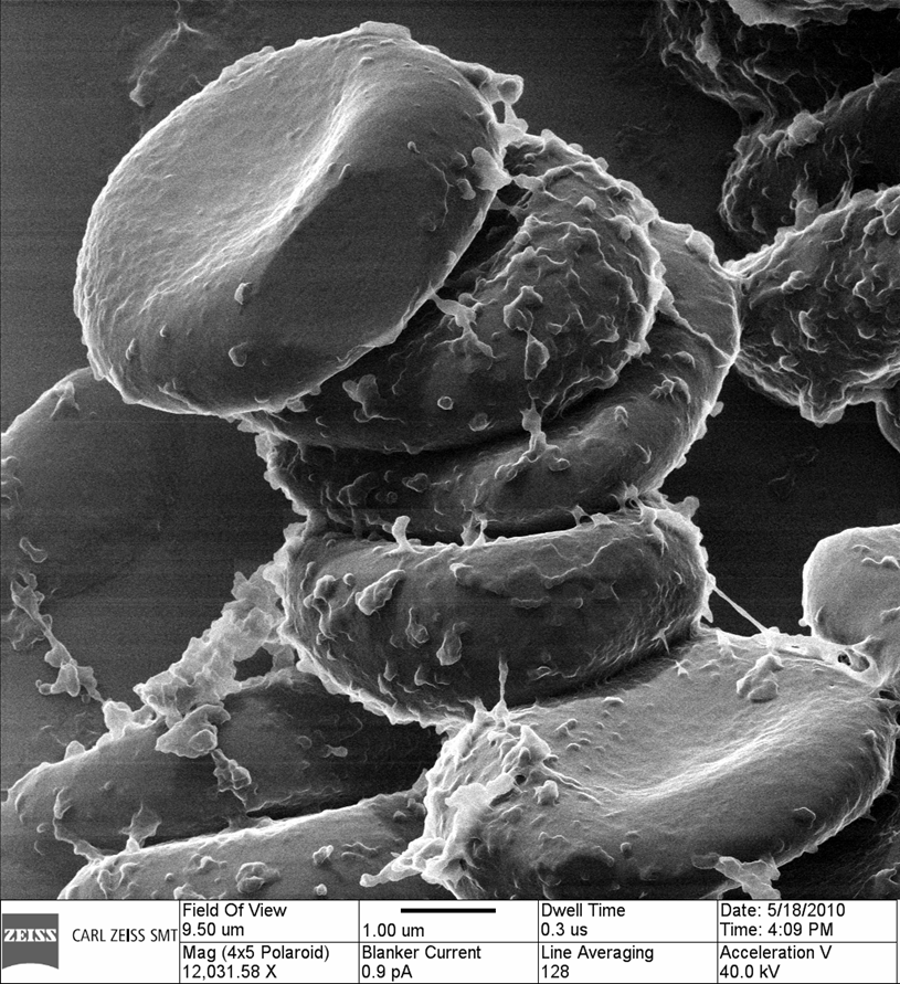
Title: REALLY short stack
Description: Platelets observed stacked up inside a blood vessel in a section of bone
Magnification (3″x4″ image): 10kX
Instrument (Make and Model):
Submitted by: Larry Scipioni
Affiliation: Carl Zeiss SMT, Inc.
HONORABLE MENTION
Title: Let’s get rid of it
Description: Top-down view of a carbon-nanotube micro-pillar after failure at ~1 GPa stress.
Magnification (3″x4″ image): 45000X
Instrument (Make and Model): Hitachi S4800 SEM
Submitted by: Siddhartha Pathak and William M. Mook
Affiliation: EMPA, Switzerland
HONORABLE MENTION
Title: New micro asteroid
Description: Scouring the surface of our silicon world we detect an unknown asteroid
Magnification (3″x4″ image): 1789X
Instrument (Make and Model): FEI Quanta 3D FEG
Submitted by: V.G. Kutchoukov and P. Kruit
Affiliation: Delft University of Technology, The Netherlands
HONORABLE MENTION
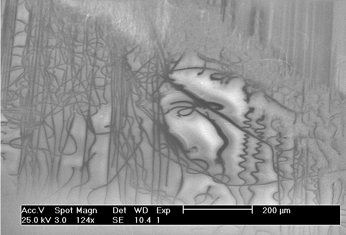
Title: Madonna Picasso
Description: A failed pattern transfer of MIM stacks from a grating mold onto a PMMA- coated glass substrate.
Magnification (3″x4″ image): 124x
Instrument (Make and Model): Instrument (Make and Model): Philips XL30 FEG SEM
Submitted by: Alex Kaplan
Affiliation: University of Michigan
HONORABLE MENTION
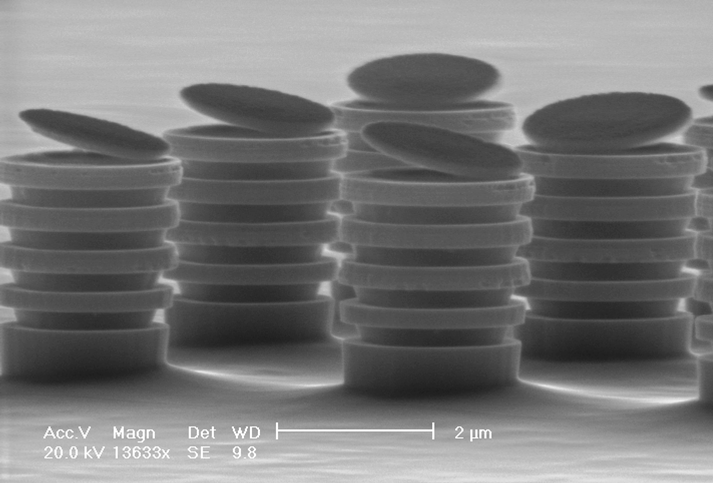
Title: Belching Trash Cans
Description: Selective etching of GaAs/AlGaAs stacks on the GaAs substrate with hard mask residue
Magnification (3″x4″ image): 13KX
Instrument (Make and Model): Philips XL30 FEG
Submitted by: Yi-Kuei Wu
Affiliation: EECS, University of Michigan, Ann Arbor
HONORABLE MENTION
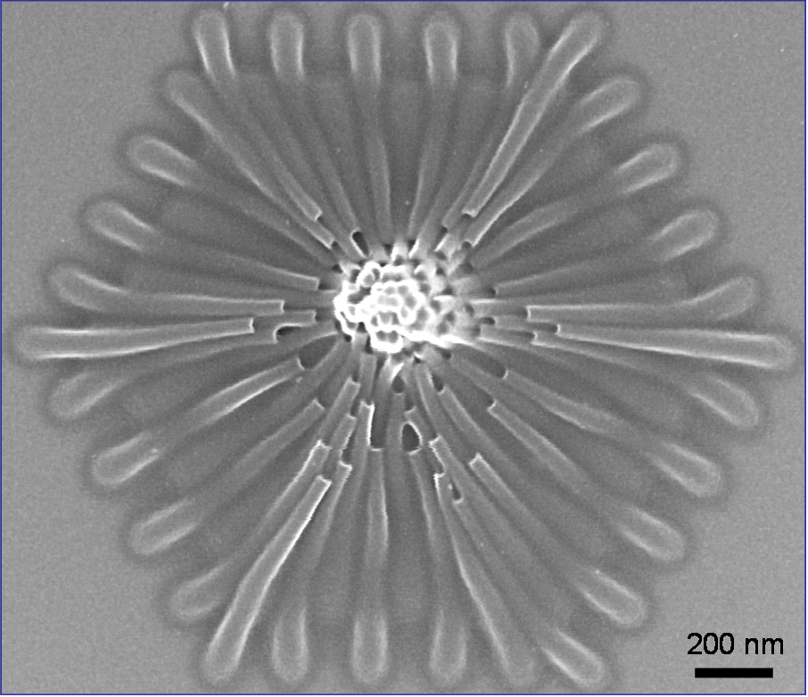
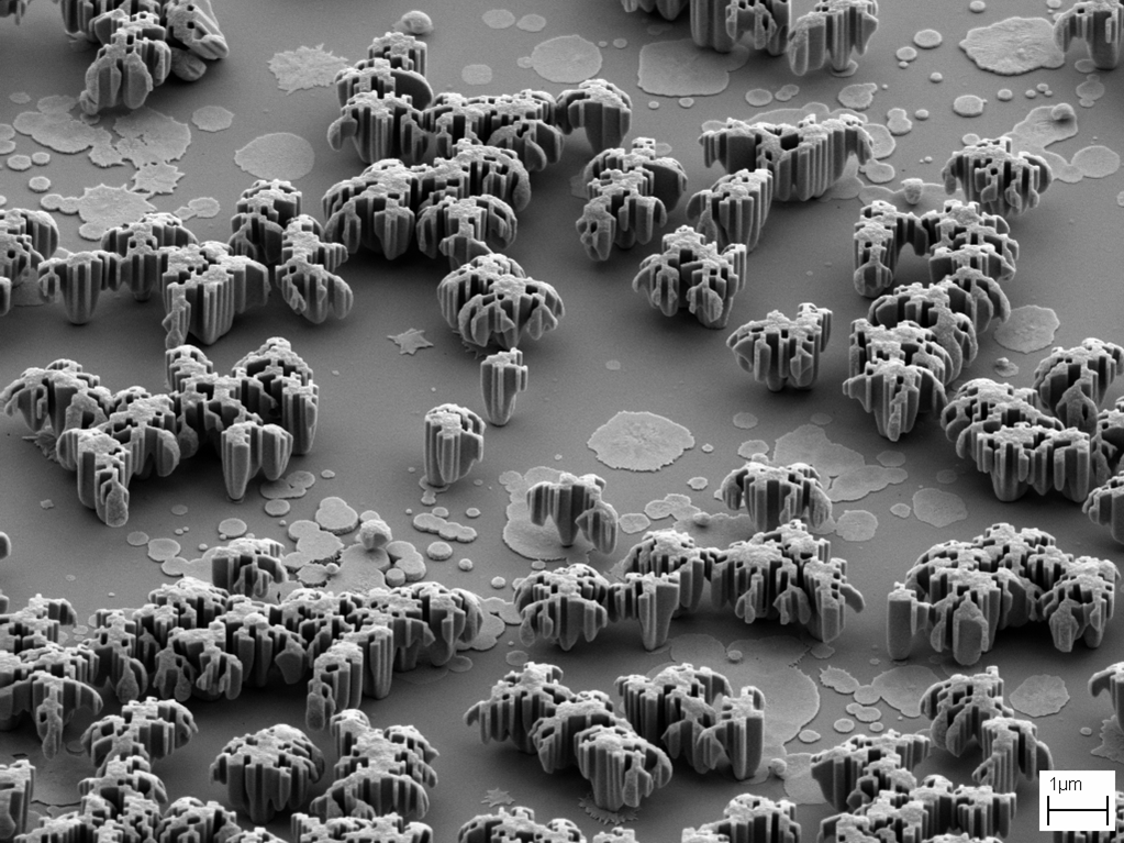
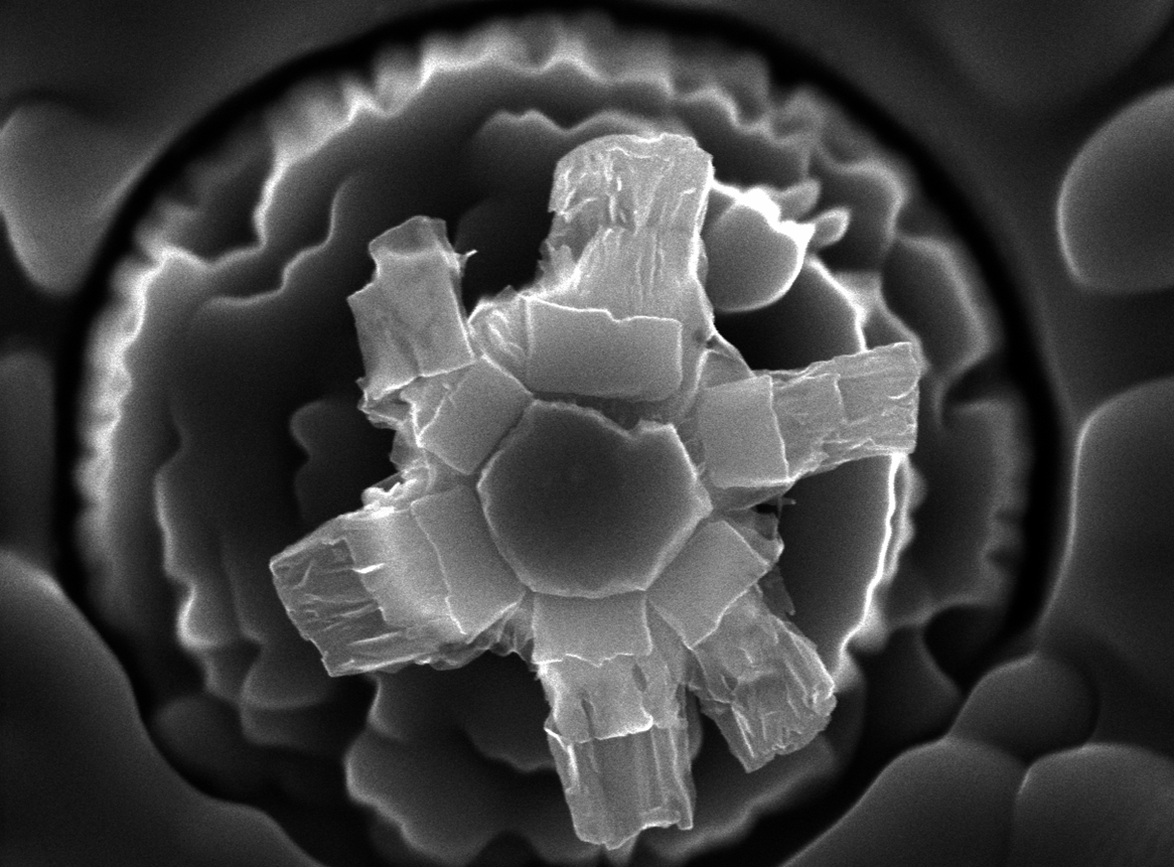
Title: “chrysanthemum”
Description:SEM image of a chrysanthemum self-assembled from electron-beam-lithography-defined PMMA nanopillars due to the capillary force during the post-development rinse and drying process. The original thickness of PMMA was ~550 nm, and PMMA was used as a negative resist. Electron-beam lithography was done by Raith 150 with an accelerating voltage of 30 kV, beam current of ~400 pA.
Magnification (5.18″x 6″ image): 75,000x
Instrument (Make and Model): Raith 150
Submitted by: Huigao Duan
Affiliation: Massachusetts Institute of Technology
HONORABLE MENTION
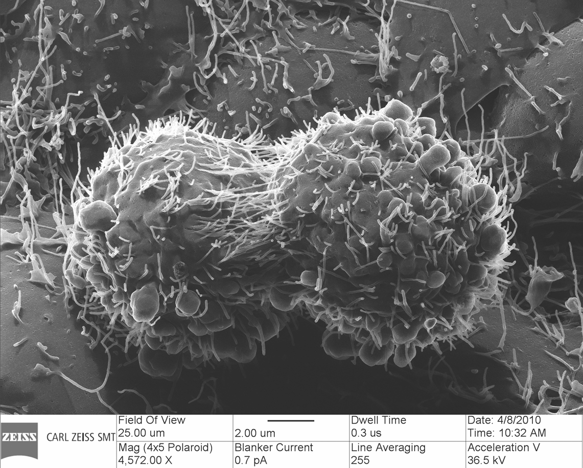
Title: Contratulations, it’s a boy
Description:These two whole cells had their micro villi entangled as if hugging.
Magnification (3″x4″ image): 4.6 kX
Instrument (Make and Model): ORION Plus He Ion Microscope
Submitted by: Shawn McVey and Dave Voci
Affiliation: Carl Zeiss SMT
HONORABLE MENTION
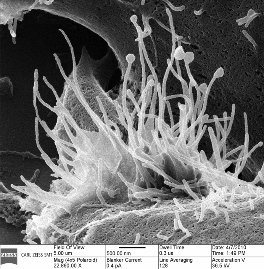
Title: Octopuses Garden
Description: This is the membrane of a mouse cell with the micro-villi reaching up – just as snakes rise for the music of the serpent charmer.
Magnification (3″x4″ image): 23 kX
Instrument (Make and Model): ORION Plus He Ion Microscope
Submitted by: Shawn McVey and Dave Voci
Affiliation: Carl Zeiss SMT
HONORABLE MENTION
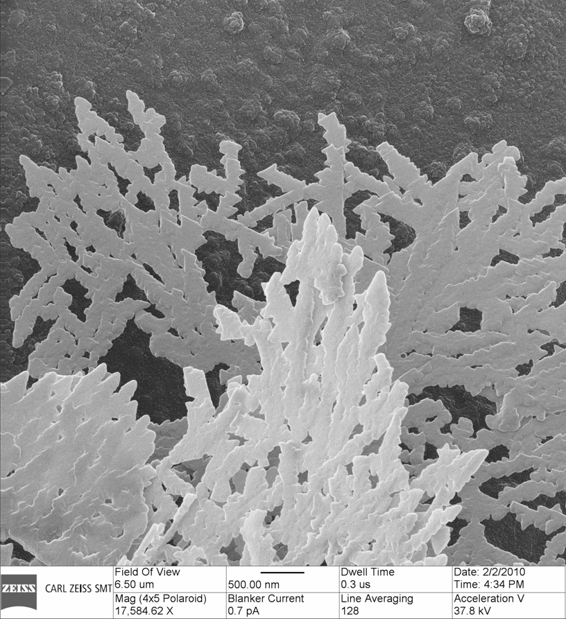
Title: Snow Flakes
Description:The planar nature of the crystal formation process is plainly visible here.
Magnification (3″x4″ image): 17kX
Instrument: Carl Zeiss, ORION Plus (He Ion Microscope)
Submitted by: Lou Farkas and Dave Voci
Affiliation: Carl Zeiss SMT
HONORABLE MENTION
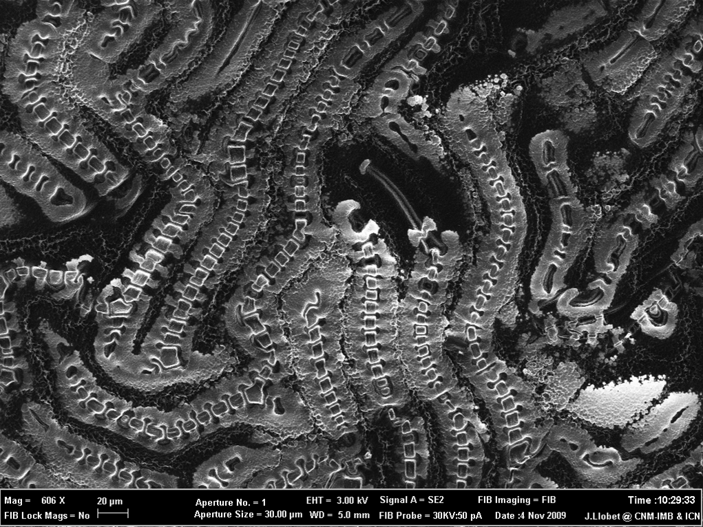
Title: I can see my house from here
Description:FIB image (Focused Ion Beam, Ga+) that shows a RIE (Reactive Ion Etching) result of an attack over an organic resist.
Magnification (3″x4″ image): 600 x
Instrument (Make and Model): CrossBeam 1560xB (Carl Zeiss)
Submitted by: Jordi Llobet1 & Aïda Varea2
Affiliation: 1IMB-CNM (CSIC) & 2 UAB – Barcelona
HONORABLE MENTION
Title: Don’t Jump!
Description:Rod of cobalt overhanging a larger silicon rod, with the “shell” formed by cobalt chloride.
Magnification (3″x4″ image): 600 x
Magnification (3″x4″ image): 29000x
Instrument (Make and Model): Zeiss Ultra55
Submitted by: Steven Hickman
Affiliation: Cornell University