←2008 |
2010→ |
The 53rd International Conference on
Electron, Ion and Photon Beam Technology
and Nanofabrication
Bizarre/Beautiful
Micrograph Contest
“ A good Micrograph is worth more than the MegaByte it consumes.”
Entries Presented by Dr. John Randall
The rules include the following:
• Entries have to be of a single image taken with a microscope and could not be significantly altered.
• There is no restriction with respect to the subject matter.
• Electron and ion micrographs have to be black and white.
In 2009, 73 entries were submitted. There were many outstanding micrographs. The work represented in the submitted micrographs covered a wide range of fields including micro mechanical, photonic, and integrated circuit fabrication, chemical and dry etching, laser optics, carbon nanotube structures, carbon nanotube growth experiments, biological samples, material science experiments and, of course, e-beam, ion beam, and photo lithography experiments.
The esteemed panel of judges who selected the award winners were:
• Don Tennant – Cornell
• Michaele Melngailis – Citizen of the World
• Tom Kenny – Stanford
There were six awards:
• Grand Prize
• Most Bizzare
• Best Photon Micrograph
• Best Ion Micrograph
• Best Electron Micrograph
• Best Video
There were 6 Honorable Mentions.
All 2009 Entries (with original titles)
Judges exercised their prerogative to liberally interpret the award categories, change the micrograph titles, and even rotate the micrograph if it pleased them.
GRAND PRIZE
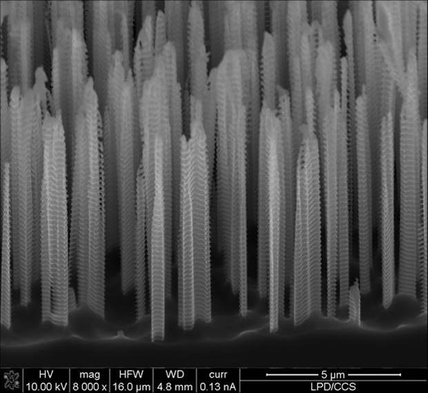
Title: Larmor City
Description:
SEM images of silicon pillar forming the black-silicon obtained by plasma etching
Magnification: (3″x4″ image): 8,000X
Instrument: FEI DB Nova 200 Scanning Electron Microscope
Submitted by: Alfredo Rodrigues Vaz, Carla Verissimo. e Clovis Fischer
Affiliation: LPD/CCS – UNICAMP – BRAZIL
Most Bizarre
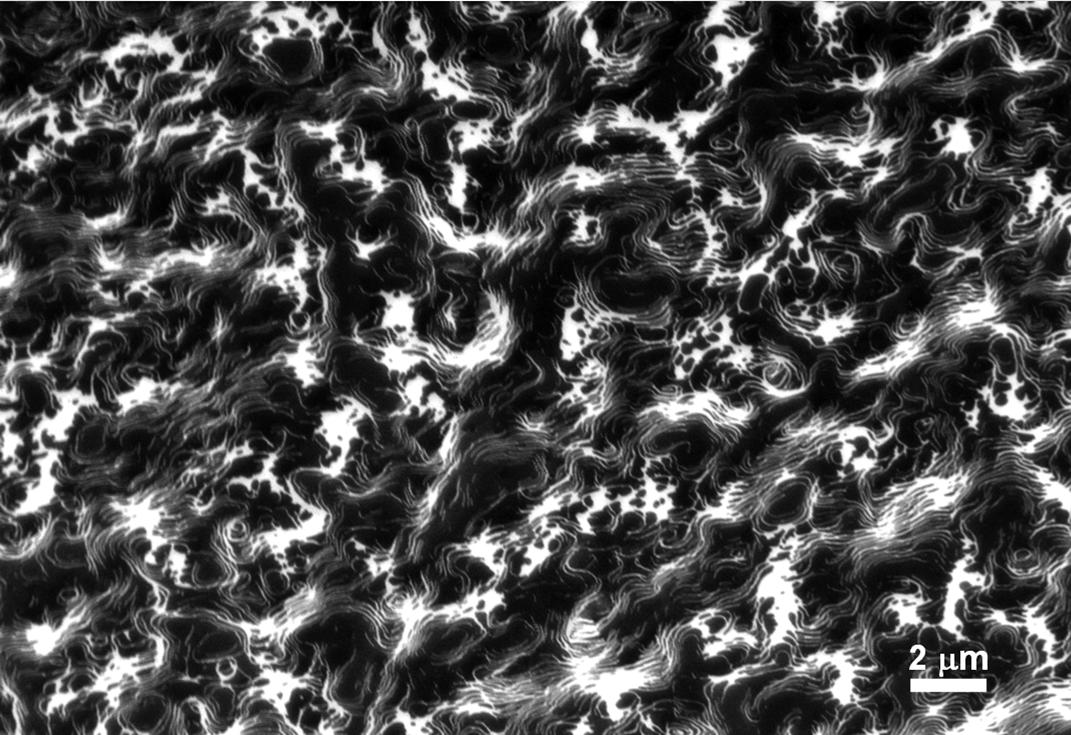
Title: Lost Souls
Description:
Rh(110) with TiOX covered regions (dark in SEM). A well-prepared Rh(110) surface with deposited titanium was heated and exposed to oxygen. The corresponding preparations and the acquisition of the depicted SEM image were conducted in an ultra-high vacuum environment. A more detailed characterization showed that the bright areas consist of pure rhodium while the dark areas are covered with titanium and oxygen.
Magnification: (3″x4″ image): 4,000X
Instrument: Omicron/Zeiss UHV Scanning Electron Microscope
Submitted by: Michael Schirmer, Marie-Madeleine Walz, Hubertus Marbach, Thomas Lukaczyk and Hans-Peter Steinruck
Affiliation: University Erlangen-Nuremberg, Germany
BEST PHOTON MICROGRAPH
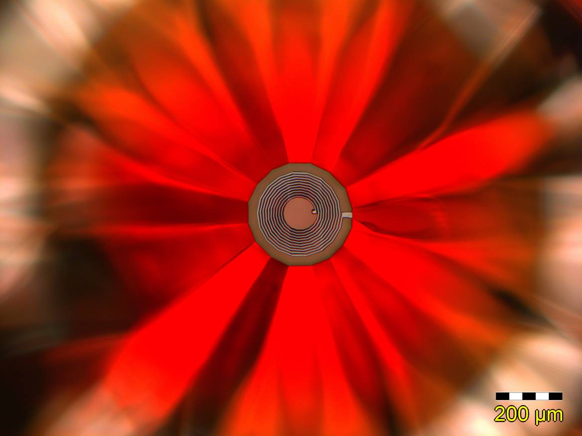
Title: Mum’s the Word
Description:
Optical micrograph of the culet of a brilliant-cut diamond that is covered with developed positive photoresist sporting the pattern of a future pick-up coil
Magnification: (3″x4″ image): 100 x
Instrument: Olympus MX-61
Submitted by: A. Imre & M. Abliz
Affiliation: Center for Nanoscale Materials, Argonne National Lab
BEST ION MICROGRAPH
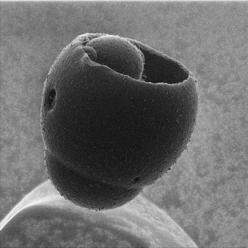
Title: The Floating Bucket of Desire
Description:
Chromatography bead mysteriously hovering above its substrate. This is an image of a core-shell chromatography bead. It is highly insulating and has no conductive coating, which may have allowed it to stack on top of an (unseen) neighbor. This particular shell has fractured, allowing the core the chance to be seen better.
Magnification: (3″x4″ image): 1,000X
Instrument: Carl Zeiss Orion Plus
Submitted by: Larry Scipioni
Affiliation: Carl Zeiss SMT, Inc.
BEST ELECTRON MICROGRAPH
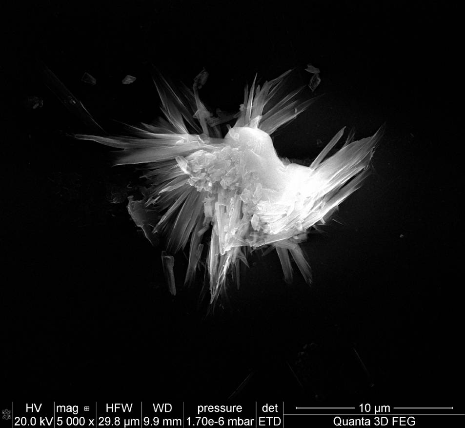
Title: “Avian Collision”
Description: Particle on aluminum on glass
I’m still looking for the micro horses… (Previous title referred to a hat and horses)
Magnification: (3″x4″ image): 5000X
Instrument: FEI Quanta 3D FEG
Submitted by: V.G. Kutchoukov and P. Kruit
Affiliation: Delft University of Technology, The Netherlands
BEST VIDEO
This Video is high quality and takes a while to download. Click link if you would like to DOWNLOAD VIDEO.
Title: Romantic Yearning
Description:
Live video (SEM imaging) of a micron-scale bridge being fabricated with a Ga+ ion beam in a dual-beam instrument by ion-beam-induced deposition (IBID) using a platinum precursor gas.
Magnification: (3″x4″ image): 25,000x
Instrument: FEI Quanta 3D FEG
Submitted by: Aurelien Botman
Affiliation: Technische Universiteit Delft (Netherlands)
HONORABLE MENTION (MicroGraph Rotated by Judges)
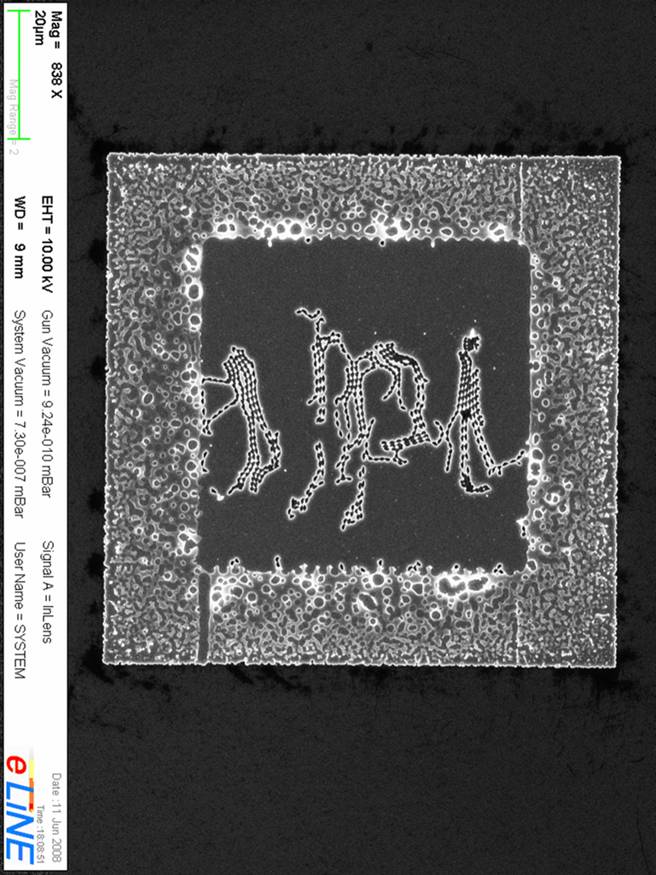
Title: Egyptian Coaster
Description:
300nm thick Al on Si after lift-off. Obviously something went terribly wrong in this e-beam exposure, but I think it’s beautiful.
Magnification: (3″x4″ image): 68.3KX
Instrument: Homemade, Raith e_line
Submitted by: Yigal Lilach & Hadar Steinberg
Affiliation: The Hebrew university of Jerusalem, Israel.
HONORABLE MENTION
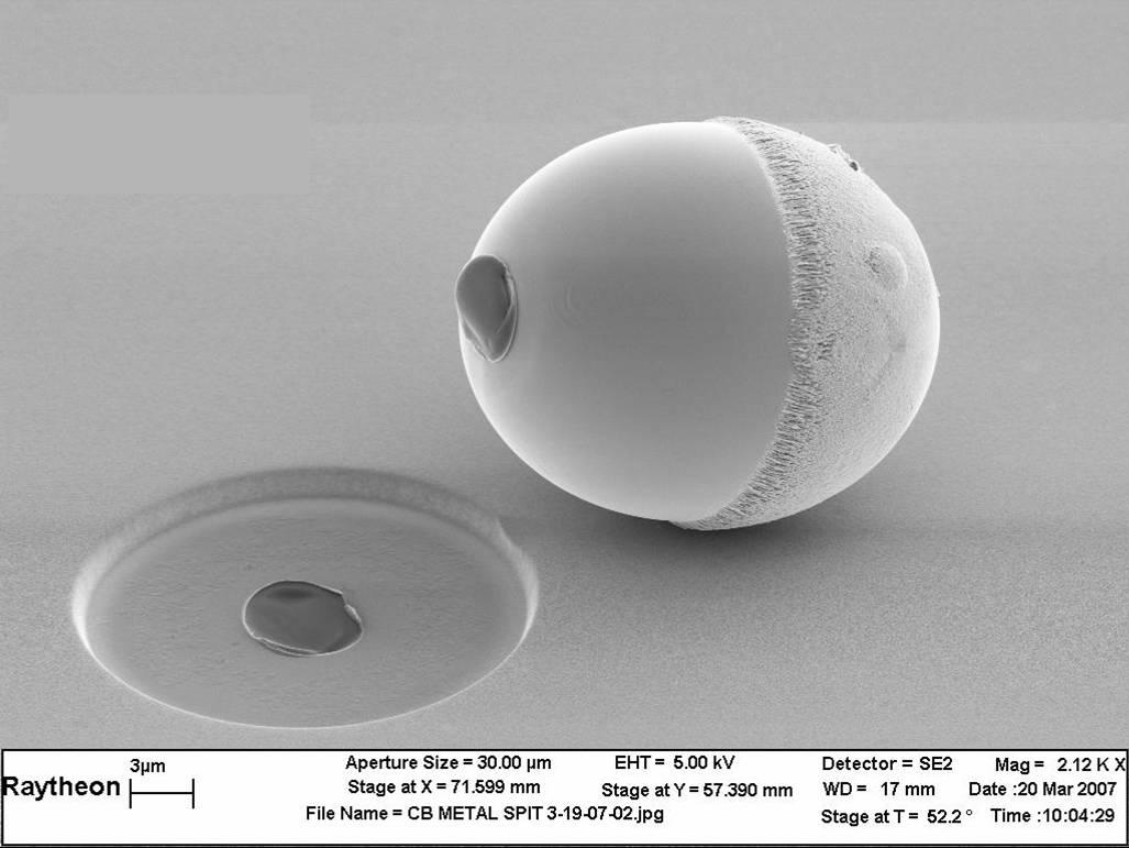
Title: Humpty Dumpty
Description:
Evaporated metal “spit”
Magnification: 2.12 KX
Instrument: Leo Genesis 1560
Submitted by: J. Pagliuca
Affiliation: Raytheon RF Components
HONORABLE MENTION
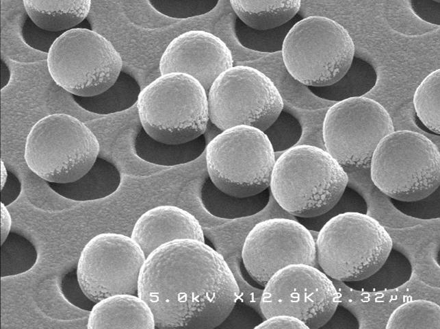
Title: “Mexican Wedding Cakes.”
Description:
Metal deposited on polystyrene microspheres leaves a culinary impression..
Magnification: (3″x4″ image): 13 KX
Instrument: LEO 1550 VP FESEM
Submitted by: Evan Brown
Affiliation: CNM – California Institute of Technology
HONORABLE MENTION
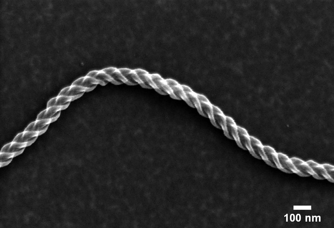
Title: “Rope a Dope”
Description:
Interwoven structure generated by CVD with ethyne resulting in the depicted “rope-shape” We acknowledge the fabrication of the shown structure by Prof. Dr. Nadejda Popovska and Katya Danova (University Erlangen-Nuremberg, Germany). Interwoven structure generated by CVDwith ethyne resulting in the depicted “rope-shape”.
Magnification: (3″x4″ image): 60.18 KX
Instrument: Omicron/ZEISS UHV SEM
Submitted by: Michael Schirmer, Marie-Madeleine Walz and Hubertus Marbach
Affiliation: University Erlangen-Nuremberg, Germany
HONORABLE MENTION
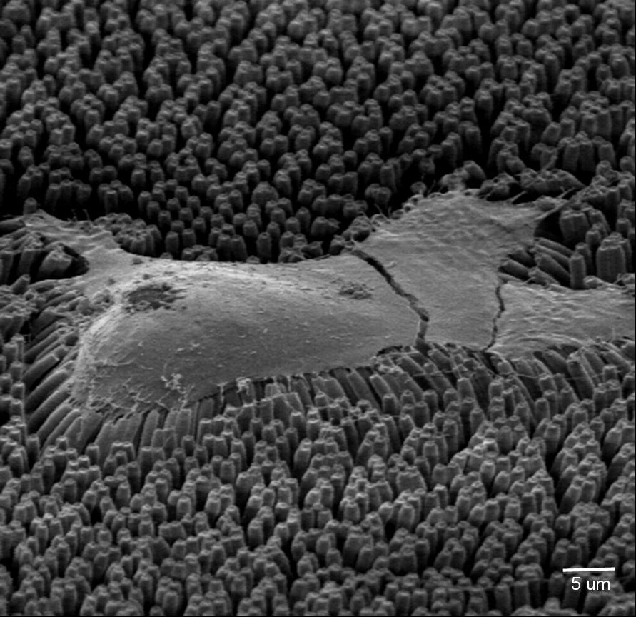
Title: Gulliver in Lilliput (the only title the judges did not change)
Description:
A 3T3 cell attached to an array of PDMS pillars. The cell on the substrate is critical point dried and gold-coated for SEM imaging. This is a SEM image of a 3T3 cell attached to the top of a hexagonal array of PDMS pillars. The cell on the substrate is critical point dried and gold-coated for SEM imaging.
Magnification:1,000X
Instrument: Hitachi 800 SEM
Submitted by: Saba Ghassemi
Affiliation: Columbia University
HONORABLE MENTION MicroGraph
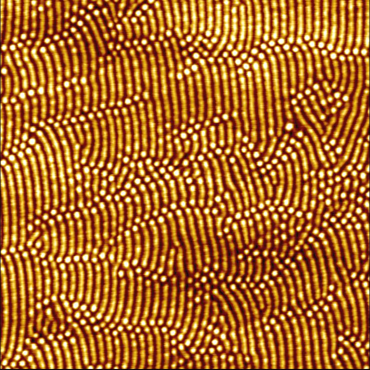
Title: A Thousand Points of Light?
Description:
Self-organized nanostructures on a Si surface induced by low-energy ion beam erosion AFM image: 2 µm x 2 µm, z-scale 3 nm
Magnification: 50 kx
Instrument: MFD-3D AFM, Asylum Research
Submitted by: J. Völlner, B. Ziberi, F. Frost
Affiliation: Leibniz-Institute of Surface Modification, Leipzig, Germany