
←2005 |
2007→ |
MNE 2006 Micro Nano Graph Contest
“ A good Micrograph is worth more than the MegaByte it consumes.”
Entries Presented by Dr. John Randall – Zyvex Labs
The rules include the following:
• Entries have to be of a single image taken with a microscope and not significantly altered.
• There is no restriction with respect to the subject matter.
• Electron and ion micrographs have to be black and white.
In 2006, 82 entries were submitted. There were many outstanding micrographs. The work represented in the submitted micrographs covered a wide range of fields including micro mechanical, photonic, and integrated circuit fabrication, chemical and dry etching, laser optics, carbon nanotube structures, carbon nanotube growth experiments, biological samples, material science experiments and, of course, e-beam, ion beam, and nano imprint lithography experiments.
The panel of judges who selected the award winners were:
- Jun-ichi Fujita – University of Tsukuba, Japan
• Lars Montelius – Lund University, Sweden
• Alex Liddle – NIST, USA
The Awards are:
• First Prize
• Second Prize
• Third Prize
The judges also selected 7 Honorable Mentions.
All 2006 Entries (with original titles)
Judges exercised their prerogative to change the micrograph titles if it pleased them.
First Prize
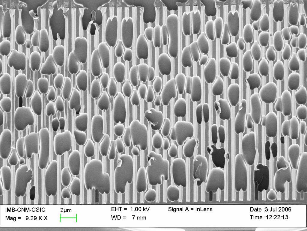
Title: Tulipes
Description:
Polymeric pattern, after NanoImprint lithography, with high imprinting and demolding temperature
Magnification (3″x4″ image): 9.29 k X
Instrument: SEM LEO 1530
Submitted by: Irene Fernández
Affiliation: CNM, IMB – Barcelona
Second Prize
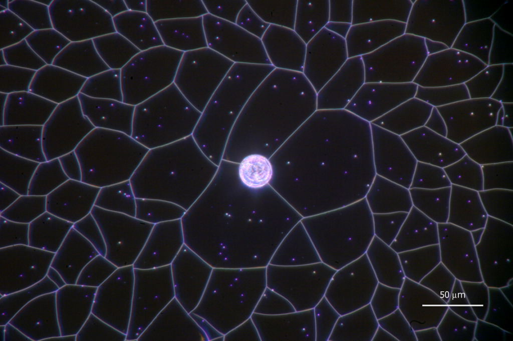
Title: “Earth caught in a Spiderweb”
Description:
A high stress nitride film crazes after laser spot crystallization of a silicon film below. Almost every vertex has no more or less than three lines emanating from it.
Magnification (3″x4″ image): 300 X
Instrument: In-situ microscope with Mitutoyo 50x objective
Submitted by: Daniel Witte
Affiliation: Stanford University, Stanford, California USA
Third Prize
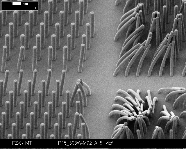
Title: “” micro-Viagra® test arrays
Description:
X-ray lithography of a 10 µm SU-8 film, fields of columns with different diameter and pitch. Patterns as indicators for the limit of stability.
Magnification (3″x4″ image): 1700 X
Instrument: M. MIKRONA SEM 525 M (Philips)
Submitted by: Timo Mappes
Affiliation: Forschungszentrum Karlsruhe GmbH, Germany
Honorable Mention
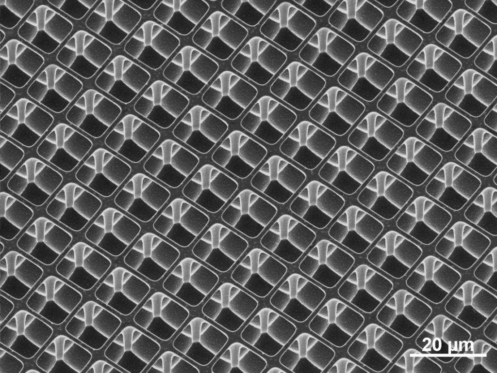
Title: Not an Escher
Description:
Accidental undercutting of square pits, during cryo etch in Si, resulting in a freestanding cage structure
Magnification (3″x4″ image): 800X
Instrument: FEI XL30 SFEG
Submitted by: Chris Rétif
Affiliation: FOM-AMOLF, Amsterdam The Netherlands
Honorable Mention
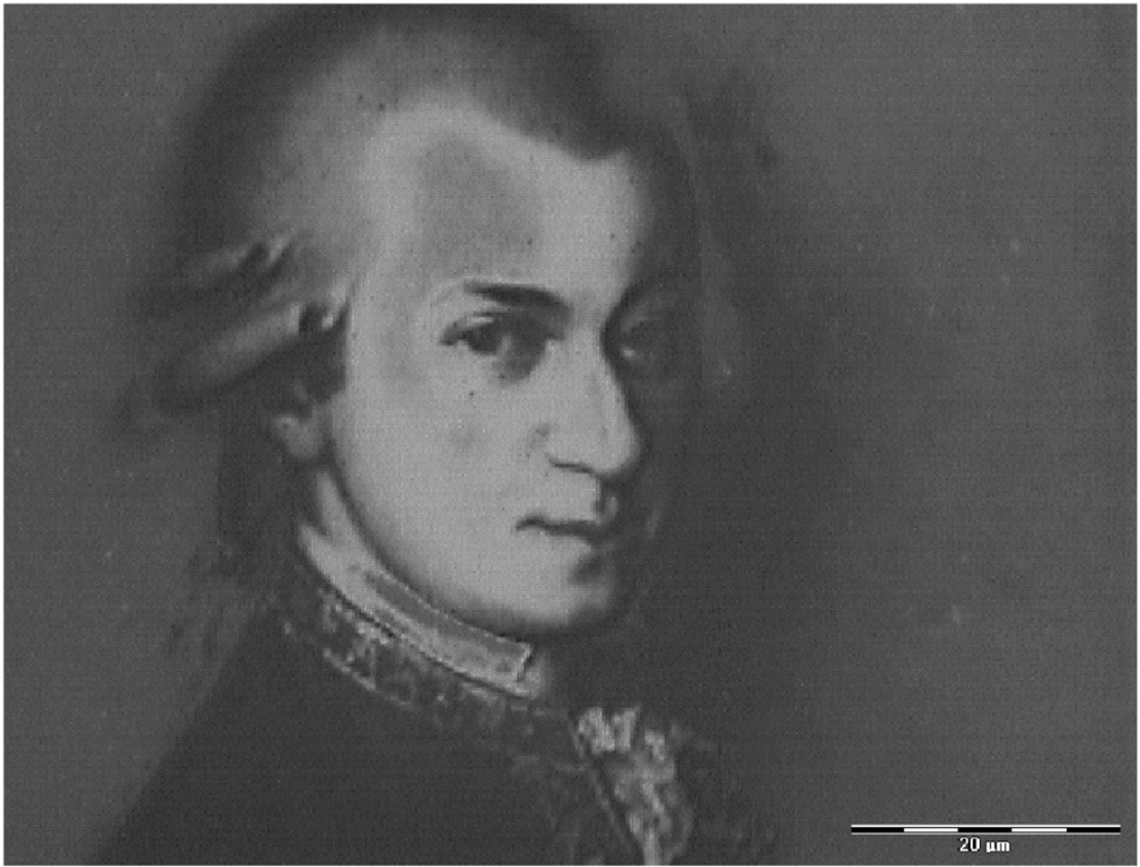
Title: Salieri’s revenge!
Description: Result of printing a real 3D structure in a UV-NIL process. Greyscale level represents depth of the structure.
Magnification (3″x4″ image): 1200 X
Instrument: LEICA INM 100 (optical microscope)
Submitted by: Guido Piaszenski
Affiliation: Raith GmbH, Germany
Honorable Mention
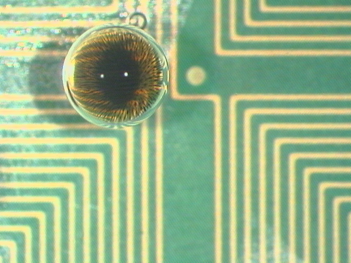
Title: Eye of Sauron
Description:
A magnetically actuated 1 ul water droplet containing super-paramagnetic particles of 250 nm diameter over a Teflon covered multilayer Printed Circuit Board. The droplet is submerged in silicone oil.
Magnification (3″x4″ image): 32X
Instrument: Zeiss Stemi V6
Submitted by: Ulrike Lehmann
Affiliation: EPFL, Lausanne, Switzerland
Honorable Mention
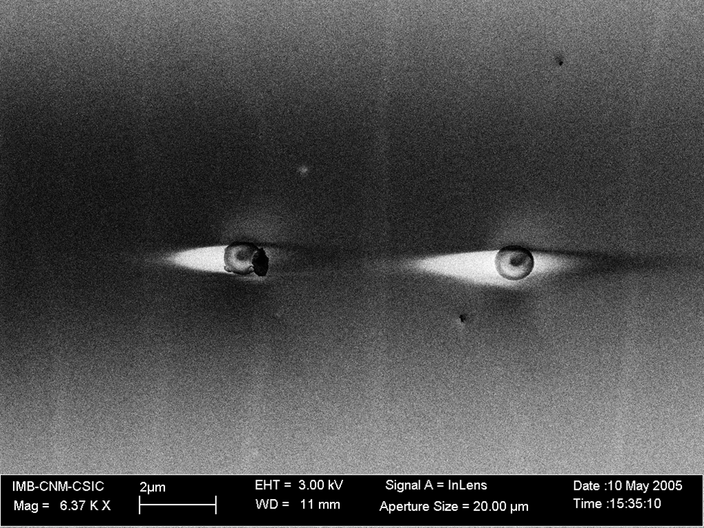
Title: Who is looking at who?
Description:
1micron latex beads onto Silicon-100 Surface
Magnification (3″x4″ image): 6.37Kx
Instrument: LEO 32
Submitted by: Jordi Teva
Affiliation: Universitat Autonoma de Barcelona
Honorable Mention
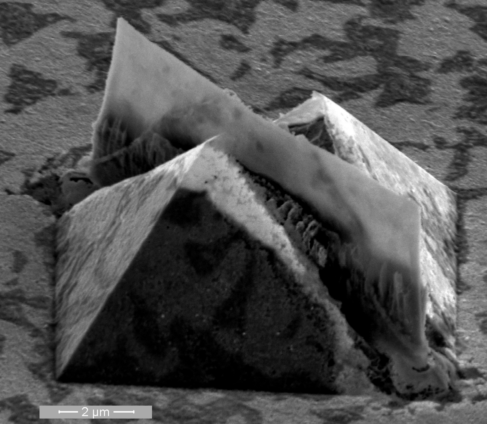
Title: Discord fell on Gizeh
Description:
Under-etched pyramid. The white material is Cu with etched Ni underneath
Magnification (3″x4″ image): 20 kX
Instrument: NOVA200 NANO SEM
Submitted by: Edouard Duriau
Affiliation: IMEC, kapeldreef 75, B3001 Leuven, Belgium
Honorable Mention
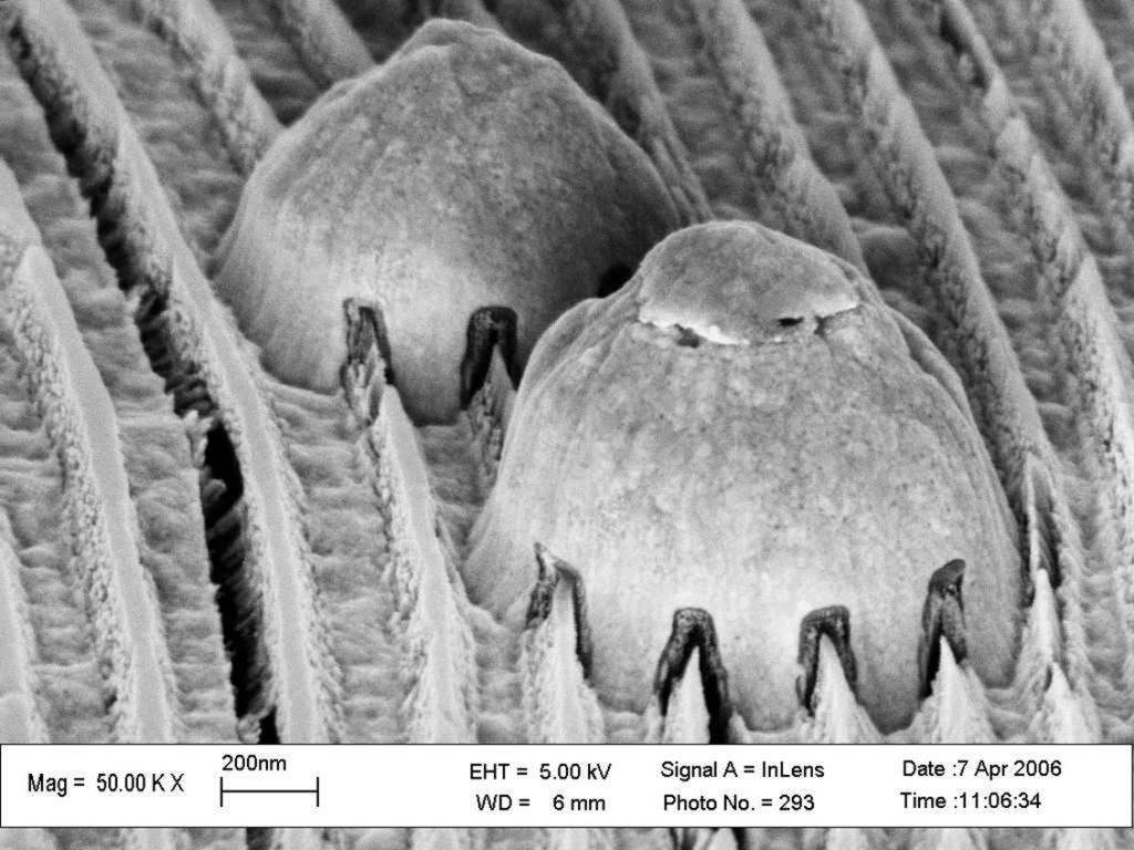
Title: Bumping Uglies
Description:
Defects on gold Fresnel zone plate after electroplating and Ar sputter etching.
Magnification (3″x4″ image): 50 KX
Instrument: ZEISS SUPRA 55VP
Submitted by:Konstantins Jefimovs
Affiliation: Laboratory for Micro- and Nanotechnology, Paul Scherrer Institut, Switzerland
Honorable Mention
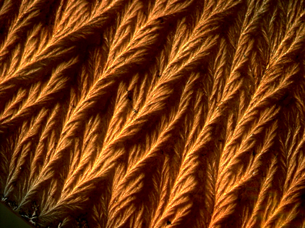
Title: Fractal Gaudi’
Description:
Optical image of quantum trees evolving from quantum dot solution by evaporation.
Magnification (3″x4″ image): Scale on the picture
Instrument: AxioCam MR
Submitted by: Yongfeng Mei
Affiliation: Max-Planck Institute for Solid State Research, Stuttgart, Germany.