
←2001 |
2003→ |
The fields of research covered by this conference have been at the forefront of the drive to develop technology to make smaller and smaller structures. We have ventured into size regimes where we are often dependent on microscopes and the skill of microscopists to see the results of our work (and often what went wrong). To highlight the importance of micrographs to the field, the conference holds a micrograph contest. The entries were judged both from the technological and artistic standpoint. Six categories were defined:
- Best Electron Micrograph
- Best Ion Micrograph
- Best Photon Micrograph
- Most Bizarre
- Best Scanning Probe Micrograph*
- Grand Prize
*The esteemed panel of judges exercised their prerogative to interpret the rules. They were not pleased with any scanning
probe micrograph that was entered and created a special prize for 2002: the “Ed Karek Award.”
The rules included the following:
- Contestants must have been registered 2002 conference attendees.
- Micrographs must be submitted as an 8 inch by 10 inch foil and must be accompanied by a completed entry sheet.
- Entries must be of a single image taken with a microscope and may not be significantly altered.
- There is no restriction with respect to the subject matter.
- Electron and ion micrographs must be black and white.
In 2002, 35 entries were submitted. There were many outstanding micrographs. The work represented in the submitted micrographs covered a wide range of fields including micro mechanical, photonic, and integrated circuit fabrication, chemical and dry etching, field emission tips, UV optics, liquid metal ion sources, metal alloy experiments, genetic experiments, carbon nanotube structures, biological samples, material science experiments, and of course e-beam, ion beam, x-ray, and photo lithography experiments. The panel of judges who selected the award winners consisted of:
Prof. Evelyn Hu
University of California at Santa Barbara
Prof. Stella Pang
University of Michigan
Don Tennant
Bell Laboratories
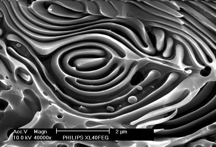
TITLE: Zebra’s Eye
Description:The micrograph is taken from a cross-section of a multiplayer structure of two amorphous polymers, poly-methylmethacrylate (PMMA) and polystyrene (PS). Such multilayers are obtained by co-extrusion of the two materials in a special mixing channel. The displayed structure is actually an unintended perturbation of the regular layer build up. The contrast is obtained by etching with O2 plasma with PMMA having a higher etchrate than PS.
Magnification for 3″x4″ image 40,000X
Instrument: Philips XL40 FEG SEM
Submitted by: Frans Holthuysen and Falco van Delft, Philips Research
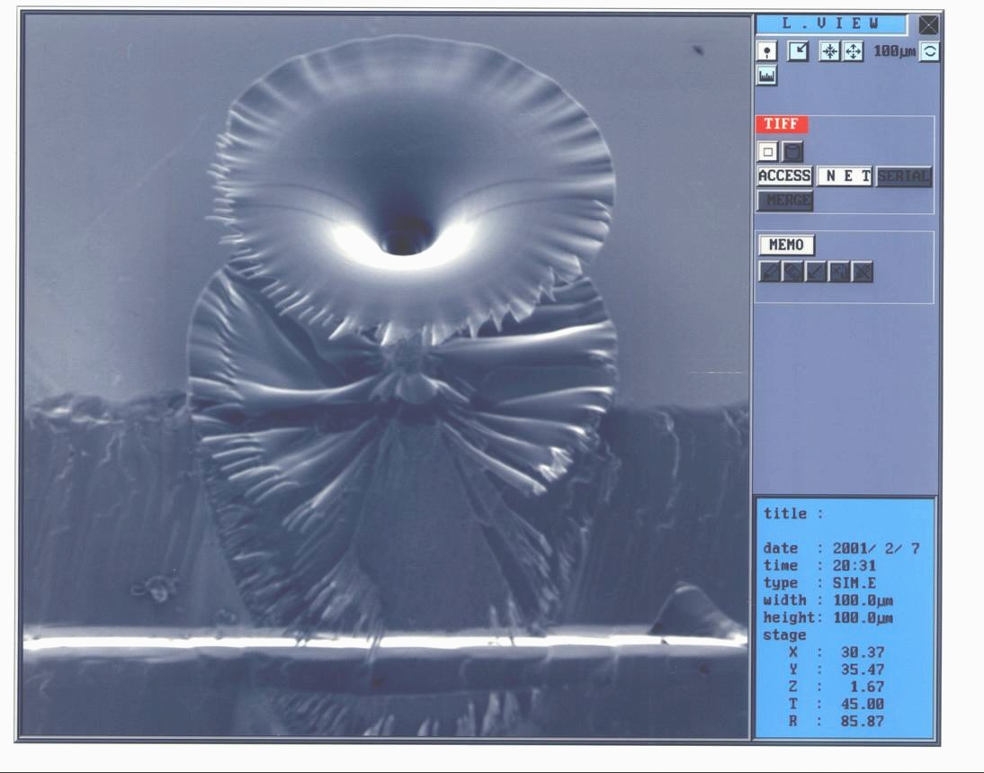
TITLE:A Morning Glory
Description: This flower was grown by 30 kV Ga focused ion beam CVD using phenanthrene gas.
Magnification 1100X
Instrument:Seiko Instruments, Inc. SMI9200
Submitted by: Shinji Matsui and Takashi Kaito, Himeji Institute of Technology
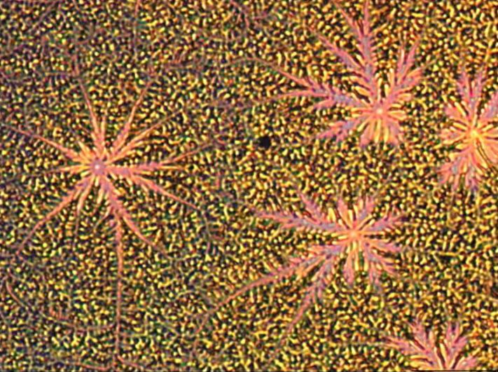
This is Your Brain…
This is Your Brain on Hydrogen Silsosquioxane. Any Questions?
TITLE: This is Your Brain… This is Your Brain on Hydrogen Silsosquioxane. Any Questions?
Description:Cracks in spin-on-glass on Si formed by incorrect thermal curing cycle.
Magnification 100X
Instrument: Nikon IC-88 Confocal (Optical) Microscope
Submitted by: Mike Fritze, MITLL

TITLE: Miss Haversham’s Wedding or Not So Great Expectations
Description: Silicon was etched to form whiskers; these structures are due to salt deposits from the etching bath.
Magnification: 8000X
Instrument: Philips XL40 FEG SEM
Submitted by: Frans Holthuysen and Falco van Delft, Philips Research
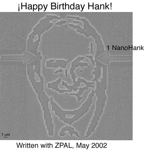
TITLE: 1 NanoHank
Description: Zpal produced image of Hank Smith at 65.
Instrument: MIT-developed Zone Plate Array Lithography System
Submitted by: Dario Gil, M.I.T.
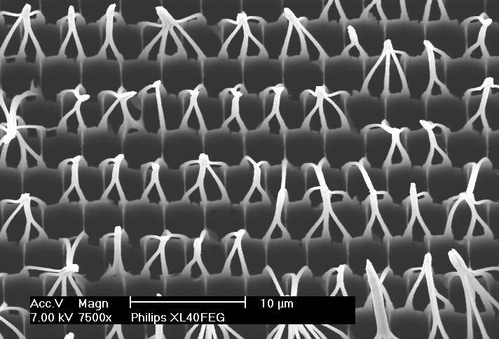
TITLE: Gumbys at the Planetarium
Description: Freestanding single-crystalline silicon whiskers with a length of 100 micrometer and a diameter of 300 nanometer are formed by etching a silicon wafer in several steps. Due to capillary forces and van der Waals interactions the wires are clustered.
Magnification for 3″x4″ image: 7500X
Instrument: Philips XL40FEG
Submitted by: Frans Holthuysen and Falco van Delft, Philips Research