←2000 |
2002→ |
The fields of research covered by this conference have been at the forefront of the drive to develop technology to make smaller and smaller structures. We have ventured into size regimes where we are often dependent on microscopes and the skill of microscopists to see the results of our work (and often what went wrong). To highlight the importance of micrographs to the field, the conference holds a micrograph contest. The entries were judged both from the technological and artistic standpoint. Six categories were defined:
- Best Electron Micrograph
- Best *Ion Micrograph
- Best Photon Micrograph
- Best *Scanning Probe MicroGraph
- Most Bizarre
- Grand Prize
*The esteemed panel of judges excersized their prerogative to interpret the rules. They were not pleased with any ion or scanning probe micrograph that was entered and awarded these prizes to electron micrographs instead.
The rules included the following:
- Contestants must have been registered 2001 conference attendees.
- Micrographs must be submitted as an 8 inch by 10 inch foil and must be accompanied by a completed entry sheet.
- Entries must be of a single image taken with a microscope and may not be significantly altered.
- There is no restriction with respect to the subject matter.
- Electron and ion micrographs must be black and white.
In 2001, 38 entries were submitted. There were many outstanding micrographs. The work represented in the submitted micrographs covered a wide range of fields including micro mechanical, photonic, and integrated circuit fabrication, chemical and dry etching, field emission tips, UV and x-ray optics, and of course e-beam, ion beam, x-ray, and photo lithography experiments. The panel of judges who selected the award winners consisted of:
Prof. Evelyn Hu
University of California at Santa Barbara
Nikki Marrion
World Bank
Dr. Al Wagner
Humanitarian, IBM
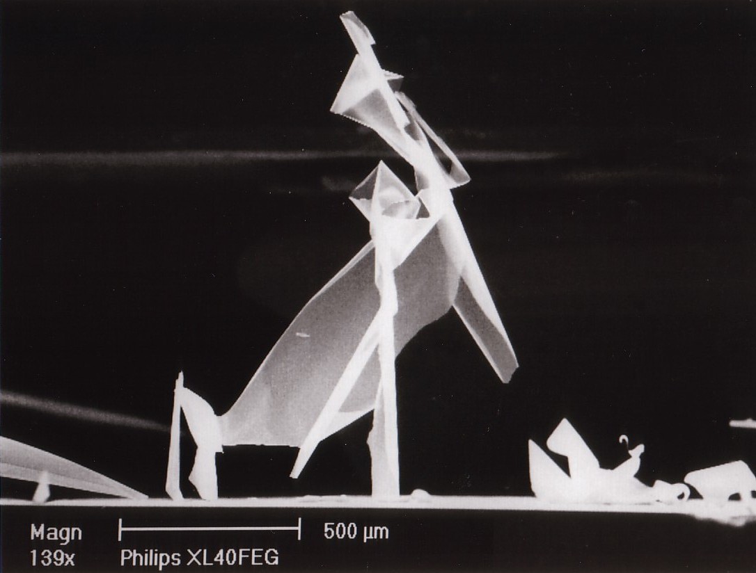
TITLE: Ballet Dancer
Description: It concerns a MgO layer deposited via spin coating of a precursor solution of Mg(OEt)2 in ethanol followed by a subsequent firing step.
Magnification for 3″x4″ image 139X
Instrument: Philips XL40 FEG SEM
Submitted by: Frans Holthuysen & Frank Dirne Philips Research Laboratories, Eindhoven, the Netherlands.
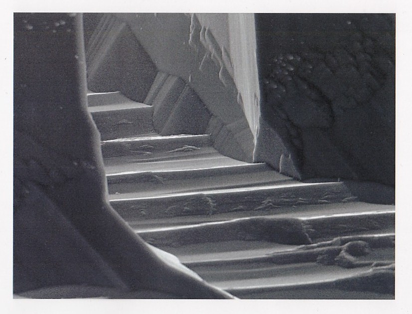
TITLE: Staircase to the Dragon’s Lair
Description: The pattern was carved unintentionally by aqueous KOH solution during wet anisotropic etching of (110) Silicon.
Magnification 1100X
Instrument: Cambridge S360 Scanning Electron Microscope* (The Judges awarded this prize in spite of the fact that this is not really an Ion Microscope.)
Submitted by: Farid Ahmed Khan Microelectronics Laboratory, University of Illinois at Urbana-Champaign
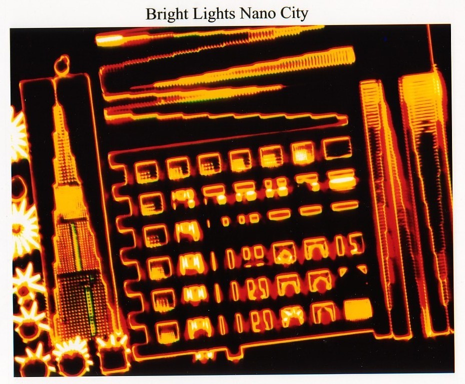
TITLE: Bright Lights Nano City
Description: This is a dark field microscope image of a test pattern used in the DEGLaSS process development. The test pattern includes a tower pattern and a variety of suspended nanostructures including 100nm-wide wires, beams, paddles, cantilevers, membranes and meshes. They are fabricated from a sing’e layer FOx SOG using dual energy (1keV and 3keV) ebeam exposures. The developed resist pattern becomes the structure without etching. The SOG can be annealed to a dense glass.
Magnification 750X
Instrument: Zeiss Axiotron
Submitted by: David M. Tanenbaum, Pomona College / Cornell University
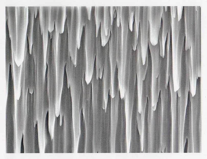
TITLE: Nano Stalactites
Description: The pattern was carved into SiC by an ICP-RIE SF6/O2 plasma when the mask got eroded during a via-hole etching attempt.
Magnification 2800X
Instrument: Cambridge S360 Scanning Electron Microscope* (The Judges awarded this prize in spite of the fact that this is not really a Scanning Probe Microscope.)
Submitted by: Farid Ahmed Khan Microelectronics Laboratory, University of Illinois at Urbana-Champaign
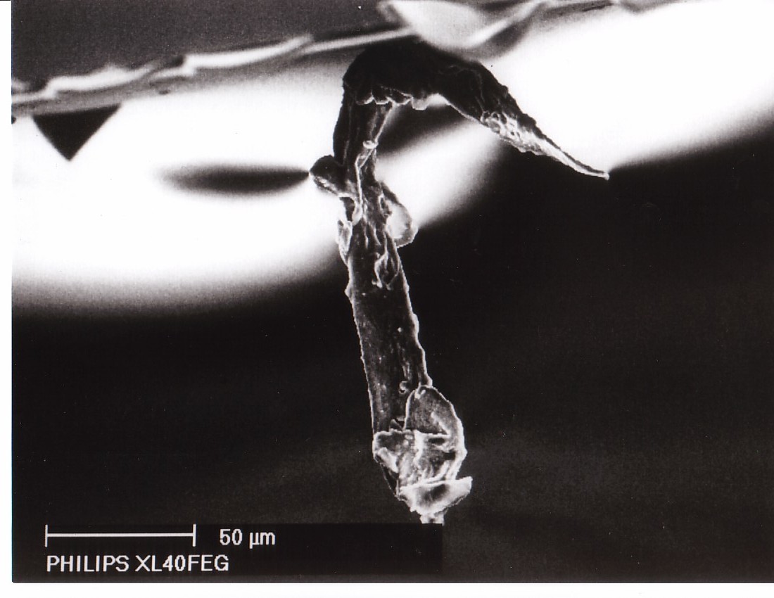
TITLE: Sea Horse
Description: The events surrounding this strange apparition are so bizarre that no one will admit to what happened.
Magnification for 3″x4″ image: 1000X
Instrument: Philips XL40 FEG SEM
Submitted by: Frans Holthuysen & Frank Dirne Philips Research Laboratories, Eindhoven, the Netherlands.
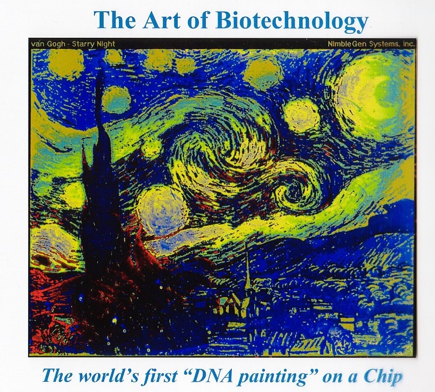
TITLE: The Art of Biotechnology
Description: A fluorescent image of a DNA chip after hybridization. False colors represent intensity of fluorescence. Chip contains 129,000 oligomers 25-basis long and was programmed from a scan of Van Gogh’s famous painting.
Magnification for 3″x4″ image: 5X
Instrument: Axon Scanner
Submitted by: Franco Cerrina, University of Wisconsin