←1997 |
1999→ |
Ion and Photon Beam Technology and NanofabricationBizarre/Beautiful Micrograph Contest
“A good Micrograph is worth more than the MegaByte it consumes.”
Results Submitted by John Randall
- Best Electron Micrograph
- Best Ion Micrograph
- Best Photon Micrograph
- Chairman’s Choice
- Most Bizarre
- Grand Prize
In addition the judges selected a number of micrographs for Honorable Mention Awards.
The rules included the following:
- Contestants must have been registered 1998 conference attendees.
- Micrographs must be submitted as an 8 inch by 10 inch and must be accompanied by a completed entry sheet.
- Entries must be of a single image taken with a microscope and may not be significantly altered.
- There is no restriction with respect to the subject matter.
- Electron and ion micrographs must be black and white.
Over 30 entries were submitted. There were many outstanding micrographs. The work represented in the submitted micrographs covered a wide range of fields including micro mechanical, photonic, and integrated circuit fabrication, chemical and dry etching, field emission tips, UV and x-ray optics, and of course e-beam, ion beam, x-ray, and photo lithography experiments. The panel of judges who selected the award winners consisted of:
Prof. Kenji Gamo
Director Gamo Lab, Osaka University
Margaret Stern
MIT Lincoln Laboratory.
Shane Palmer
Senior Member of the Techncal Staff – Texas Instruments
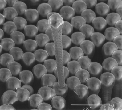
Title: And how are you today?
Magnification: 16,500X
Instrument: Hitachi Scanning Electron Microscope
Submitted by: Maggie Hupcey, Cornell University and Nancy LaBianca IBM T.J. Watson Research Center.
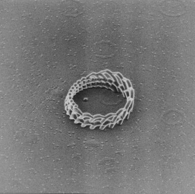
Title: Micro Collseum
Magnification (for 4″ Height):
Instrument: Seiko SMI-9800
Submitted by: S. Matsui, SELETE and T. Kaito Seiko Instruments Inc.
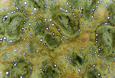
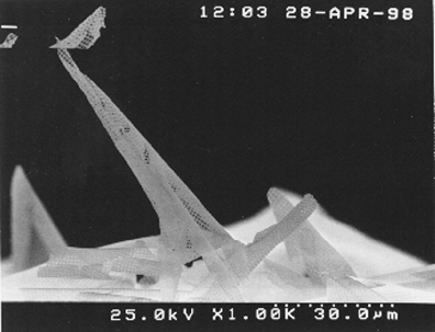
Title: Swamp Thing.
Magnification (for 4″ image height): 400X
Instrument: Nikon
Submitted by: Magie Hupcey and Yayoi Takamura – Cornell University
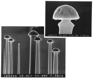
Title: No their not mushrooms.
Magnification: 4,000X/18,000X
Instrument: Hitachi – 4000 FEM
Submitted by:Ivo W. Rangelow University of Kassel, Germany
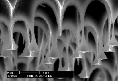
Title: Flying Alhambra
Magnification: 40,000X
Instrument: Philips XL40FEG SEM
Submitted by: Frans Holthuysen and Jos Weterings – Philips Research Laboratories, Einhoven
Title: PPMS Parakeet
Magnification (for 4″ Height): 1,000X
Instrument: Hitachi S-4500 Scanning Electron Microscope
Submitted by: Mike Nault – Applied Materials
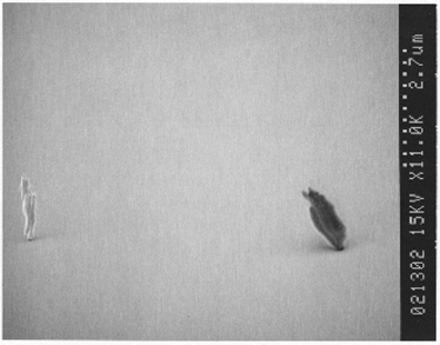
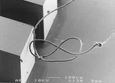
Title: There’s one in every crowd.
Magnification (for 4″ Height): 3,000X
Instrument: JEOL JSM-6400FV Scanning Electron Microscope
Submitted by: Joel Wendt and Stan Kravitz – Sandia National Laboratories
Title: The real reason for good adhesion in deep x-ray Lithography
Magnification (for 4″ Height): 130X
Instrument: Scanning Electron Microscope
Submitted by: Franz Joseph Pantenburg – Institut fur Mikrostrukturtechnik