←2011 |
2013→ |
Bizarre/Beautiful
Micrograph Contest
“A good Micrograph is worth more than the MegaByte it consumes.”
Entries Presented by Dr. John Randall
The rules include the following:
• Entries have to be of a single image taken with a microscope and could not be significantly altered.
• There is no restriction with respect to the subject matter.
• Electron and ion micrographs have to be black and white.
In 2012, 79 entries were submitted. Including:
• 45 Electron Micrographs
• 13 Ion Micrographs
• 12 Photon Micrographs
• 7 Video Micrographs
• 2 Scanning Probe Micrographs
The panel of judges who selected the award winners were:
• Judy Tennant
• Stella Pang
• ShaChelle Devlin Manning
The Judges exercised their prerogative to liberally interpret the award categories, change the micrograph titles, and even rotate the micrograph if it pleased them.
There were six awards:
• Grand Prize
• Most Bizzare
• Best Photon Micrograph
• Best Ion Micrograph
• Best Electron Micrograph
• Best Video Micrograph
There were 4 Honorable Mentions.
All 2012 Entries (with original titles)
BEST ELECTRON MICROGRAPH
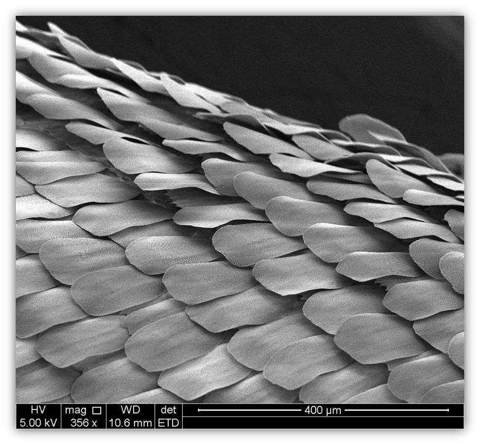
Title: Colored Chocolate
Description: The scales array on a butter fly wing.
Magnification (3″x4″ image): 356X
Instrument (Make and Model): FEI Quanta FEG
Submitted by: Fei Ding
Affiliation: Princeton University
BEST ION MICROGRAPH
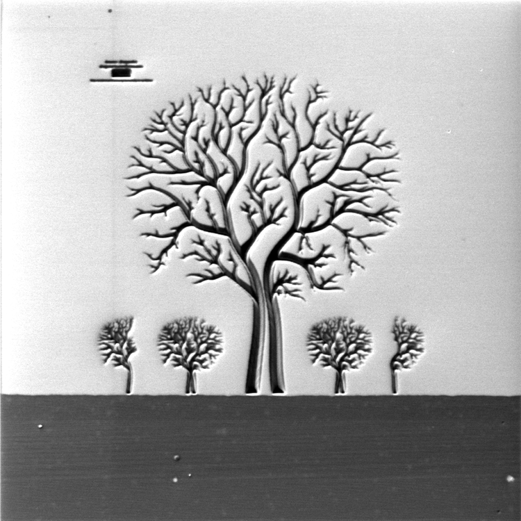
Title: THE BODHI TREE
Description: PIECE OF ART MADE BY SPUTTERING SILICON OXIDE ON SILICON USING GALLIUM FIB (UNCOATED SAMPLE)
Magnification (3″x4″ image): 5715 X
Instrument: Carl Zeiss ORION Plus
Submitted by: Mohan Ananth
Affiliation: Carl Zeiss NTS
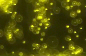
BEST PHOTON MICROGRAPH
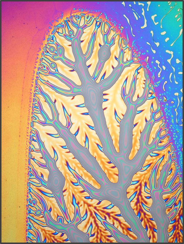
Title: The Fractal Longboard
Description: Inspired by the book Exodus. But we call him George.
Magnification: 850X (120µm x 90µm image size)
Instrument: Nikon Eclipse LV150 with DS-5M camera
Submitted by: Iris Bergmair / Lukas Häusler
Affiliation: PROFACTOR GmbH
MOST BIZARRE MICROGRAPH
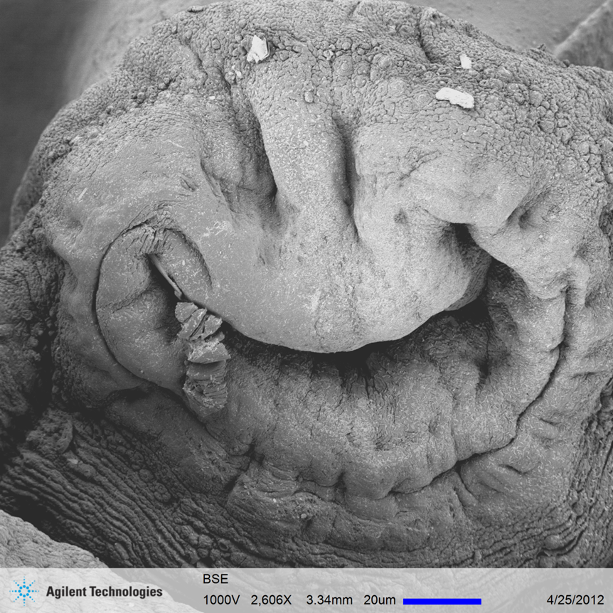
Title: Me eat cookie!
Description: The oral sucker of an adult liver fluke. (Sample courtesy of Institute of Cytology and Genetics, Novosibirsk, Russia.)
Magnification (3″x4″ image): 2,606X
Instrument: Agilent 8500 FE-SEM
Submitted by: C. Silver, L. Muray & J. Spallas
Affiliation:Agilent Technologies
BEST VIDEO MICROGRAPH
Title: Nanopecker
Description: Free standing “Nanowoodpecker”, fabricated on the edge of an SOI substrate – applying a sequential electron beam induced deposition (EBID) process. Tungsten nanoprobes for ac-field application make him move around. This is a (non-real-time) video (with sound!) showing the sequential “making of” the nanowoodpecker with EBID, using a Pt-precursor and patterning on image (POI) feature. Afterwards, tungsten tipped nanoprobers are approached to close proximity of the nanowoodpecker´s insulating SiOx reservoir, which has been deposited as well at the supporting foot pillar. During e-beam exposure while scanning, explicitely this reservoir (and most likely other parts of the nanowoodpecker as well) charge. An ac-voltage in the order of a few Volts is applied to the nanoprobers thus applying an ac-electrical field, which makes the nanowoodpecker move around due to varying electrical forces affecting its charged reservoir.
Magnification (3″x4″ image): 5000X
Instrument: Raith eLine-plus
Submitted by: F. Nouvertné, A. Rudzinski
Affiliation: Raith GmbH
GRAND PRIZE
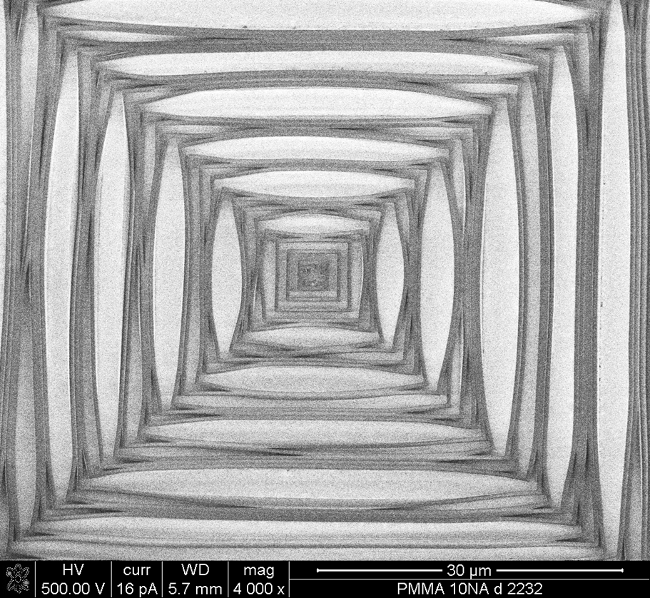
Title: Going Up or Going Down?
Description: Square box of 500nm line segments and spaces overexposed in PMMA
Magnification (3″x4″ image):4000x
Instrument: FEI Nova 600
Submitted by: Guy DeRose
Affiliation: Caltech
HONORABLE MENTION
Title: DNA Line Dancing
Description: Fluorescent, Charged, Polystyrene nanoparticles (200nm) are being actively manipulated on 40nm e-beam written lines made out of gold. Mimicking DNA-Protein interactions in an artificially created system. The video shows the 1-d diffusion and trapping/de-trapping nature of the nanoparticles under the action of electrostatic fields.
Magnification (3″x4″ image): 100x
Instrument: Nikon Ti
Submitted by: Ashwin Panday
Affiliation: L. Jay Guo, University of Michigan Japan
HONORABLE MENTION
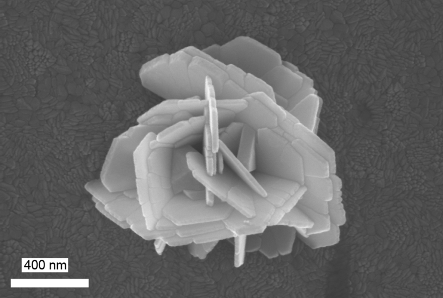
Title: Pua
Description: Rose-like particle, on glass-ITO, made of silver plates obtained by laser-induced liquid deposition. Two glass-ITO slides holding a liquid silver precursor were illuminated with an Nd:YAG laser with a 532 nm wavelength. Interestingly, the beam was not focused on the surface of the picture. It was focused on a previous slide which was melted by the powerful beam.
Magnification (3″x4″ image): 45,100X
Instrument: Raith e-Line
Submitted by: Carlos A. Jarro
Affiliation: University of Kentucky
HONORABLE MENTION
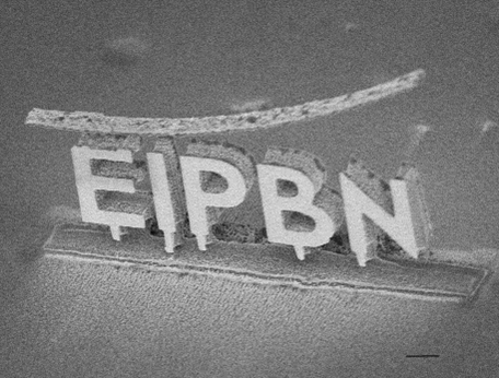
Title: Raising an eyebrow @ EIPBN
Description: This is the 3D Si structure we made as the first demonstration of our new 3D nanofabrication technique. Although the bending band on the top was not intended to put, it remained by happening and we think that it looks nice. It was used as a mask for the etching from the top at the first fabrication step, and it should be removed, we planned, at the final step of etching from the front and back sides.
Magnification (3″x4″ image):6.5KX
Instrument: Hitachi S-7800H
Submitted by: K. Yamazaki, H Yamaguchi
Affiliation: NTT Basic Research Labs.
HONORABLE MENTION
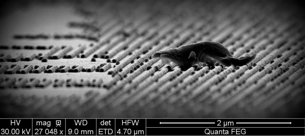
Title: Micro-NA Honey Badger
Description: A badger wanders on a nano-structured plain, weaving towards a defect river. Working distance is modified during scanning for artistic effect.
Magnification (3″x4″ image):27,048x
Instrument (Make and Model): FEI Quanta FEG
Submitted by: Fei Ding
Affiliation: Princeton University