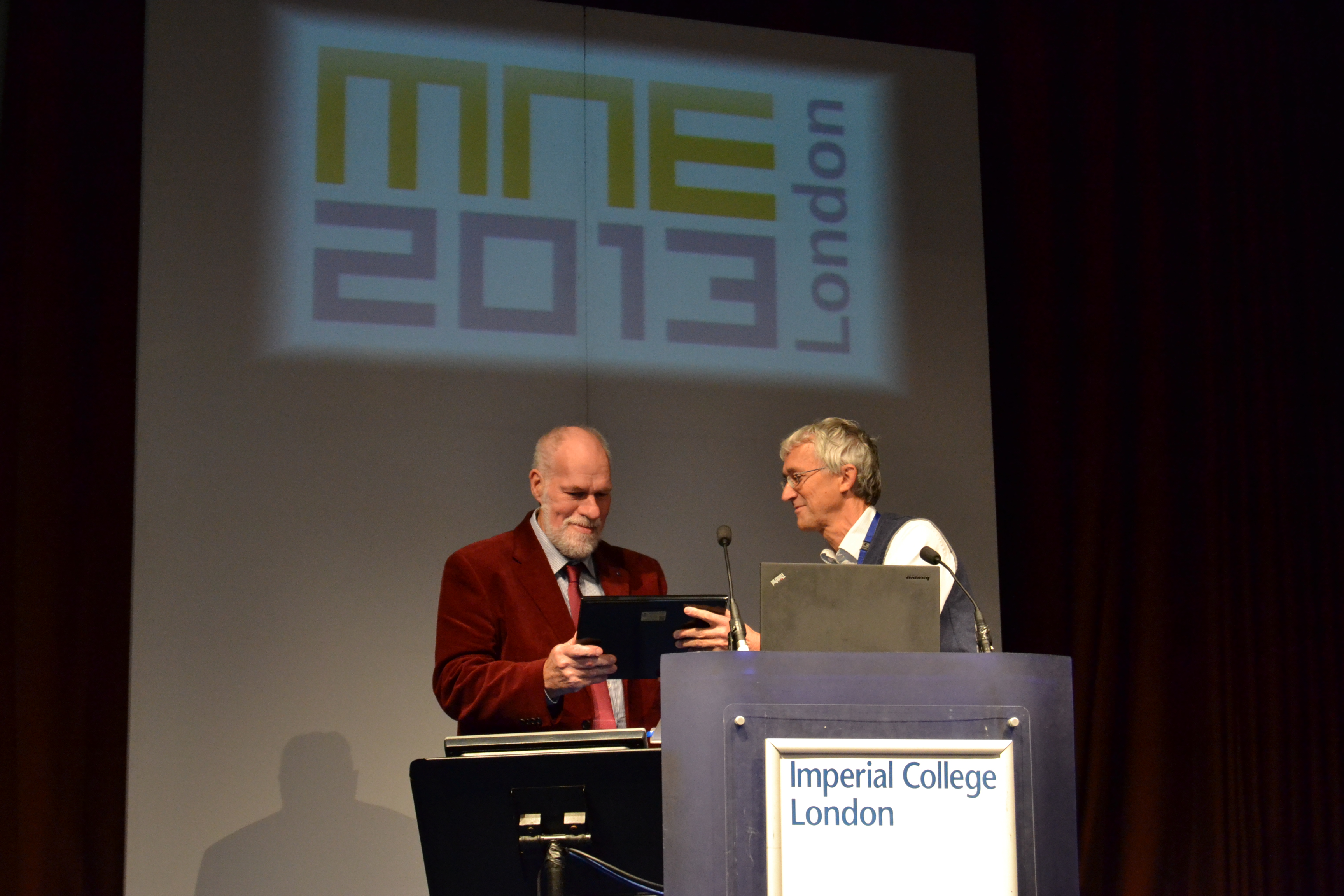
On September 19, 2013 in the Great Hall of the Imperial College of London at the Micro and Nano Engineering Conference with over 600 attendees and exhibitors, a lifetime achievement award:
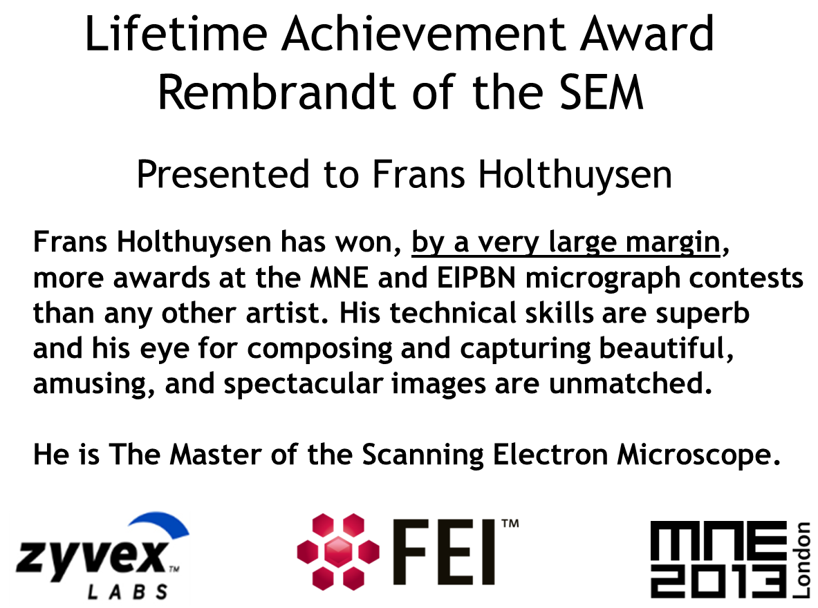
"The Rembrandt of the SEM" was given to Frans Holthuysen of Philips Research Labs.
 |
 |
| Frans receives award from FEI's Hans Mulders. |
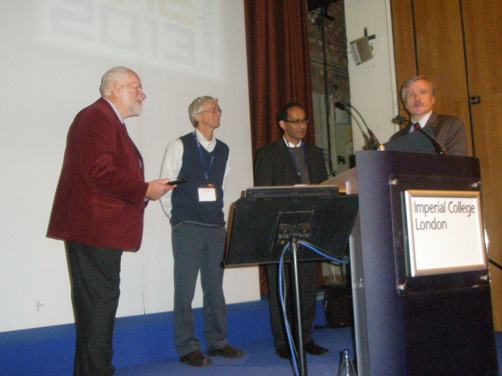
Left to right: Frans Holthuysen, Hans Mulders, Zahid Durrani, John Randall
|
The award was sponsored by FEI whose SEM was the instrument that Frans used to take most of his micrographs. An award plaque was presented by Hans Mulders of FIE a long time friend of Frans. The Plaque was inscribed as follows:
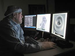
On September 25, 2013, Frans Holthuysen retired from Philips Research Labs after 48 years of service. This is a loss to Philips and to the rest of the world. His rare talent did what rare talent does, it breaks boundaries. His skill was such that it broke out of the technical world into the world of art. His work was displayed at the New York Museum of Modern Art and many other places. Those of us who have marveled at his skill and artistry will sorely miss seeing what new wonders that he has managed to capture with an SEM. However, we will have to be content with his legacy of spectacular images. A sampling is displayed below.
Just for fun type "Frans Holthuysen" into Google images and see what is displayed.

Title : ōFlying Alhambraö
Magnification (3"x4" image): 40KX
Instrument: Philips XL-40 FEG Scanning Electron Microscope

Title : ōUnder Constructionö
Magnification (3"x4" image): 3KX
Instrument: Philips XL-40 FEG Scanning Electron Microscope

Title : ōBallet Dancerö
Magnification (3"x4" image): 139X
Instrument: Philips XL-40 FEG Scanning Electron Microscope

Title : ōSea Horseö
Magnification (3"x4" image): 1000X
Instrument: Philips XL-40 FEG Scanning Electron Microscope

Title : ōZebra's Eyeö
Magnification (3"x4" image): 40KX
Instrument: Philips XL-40 FEG Scanning Electron Microscope

Title : ōMiss HavershamÆs Wedding or Not So Great Expectationsö
Magnification (3"x4" image): 8000X
Instrument: Philips XL-40 FEG Scanning Electron Microscope

Title : ōGumbys at the Planetariumö
Magnification (3"x4" image): 7500X
Instrument: Philips XL-40 FEG Scanning Electron Microscope

Title : ōDancing Girlsö
Magnification (3"x4" image): 50KX
Instrument: Philips XL-40 FEG Scanning Electron Microscope

Title : ōWisconsin Thongö
Magnification (3"x4" image): 1000X
Instrument: Philips XL-40 FEG Scanning Electron Microscope

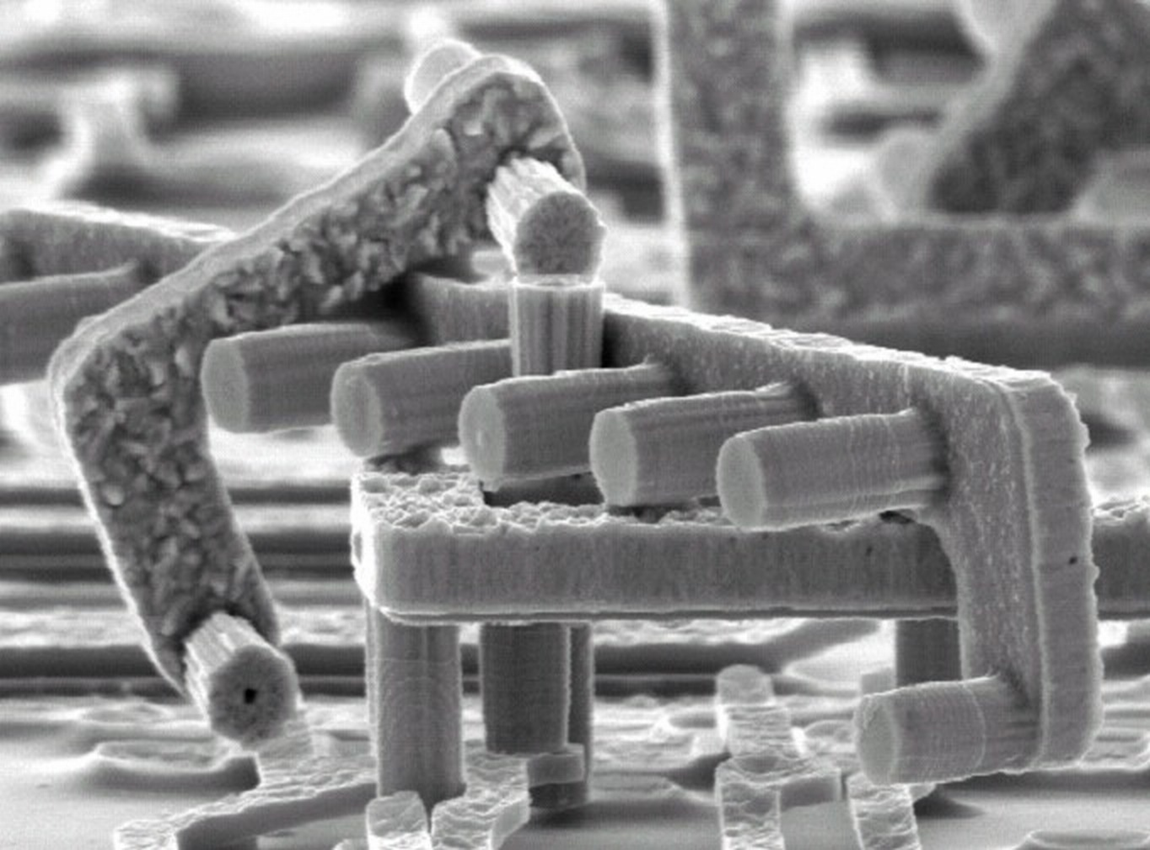
Title : ōPico Predatorö
Magnification (3"x4" image): 340X
Instrument: FEI NovaNanoSEM600

Title : ōTower of Babylonö
Magnification (3"x4" image): 60X
Instrument: FEI NovaNanoSEM600

Title : ōSpiders Websiteö
Magnification (3"x4" image): 240X
Instrument: FEI NovaNanoSEM600

Title : ōWest Side Storyö
Magnification (3"x4" image): 20KX
Instrument: Philips XL-40 FEG Scanning Electron Microscope

Title : ōFailure to Igniteö
Magnification (3"x4" image): 5,000X
Instrument: FEI NovaNanoSEM600

Title : ō Caves (Stalagnieten) ö
Magnification (3"x4" image): 4,000X
Instrument: FEI NovaNanoSEM600

Title : ō Donut ö
Magnification (3"x4" image): 1,250X
Instrument: FEI NovaNanoSEM600

Title : ō Leaves ö
Magnification (3"x4" image): 100KX
Instrument: FEI NovaNanoSEM600

Title : ōThe Waveö
Magnification (3"x4" image): 40KX
Instrument: FEI NovaNanoSEM600
Title : "The Ruins of Damascene"
Magnification (3"x4" image): 10KX
Instrument: Philips XL-40 FEG Scanning Electron Microscope
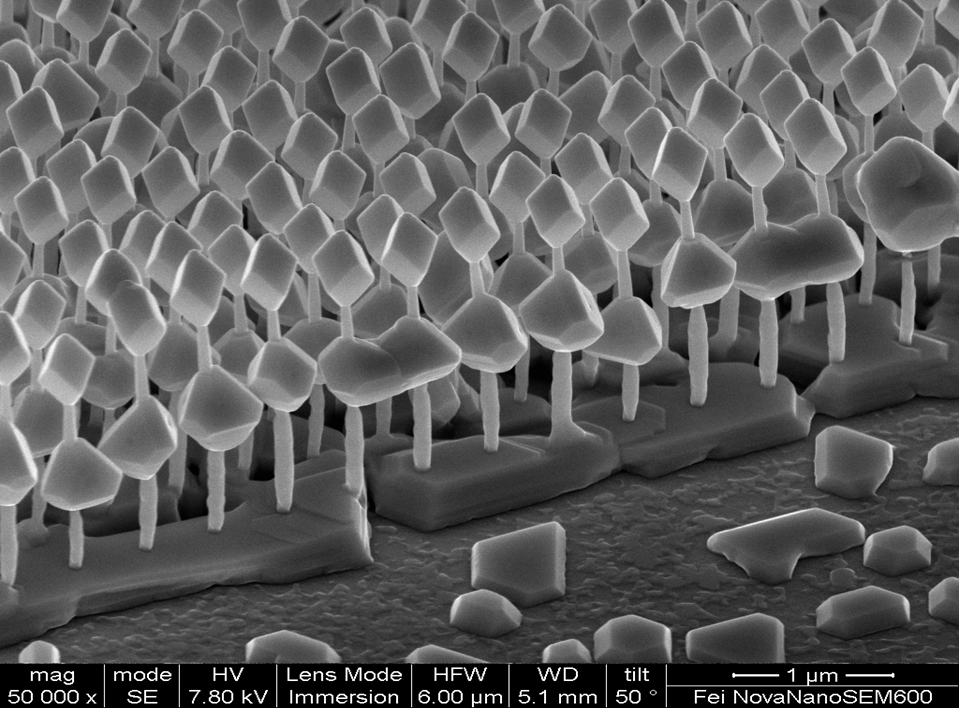
Title : ōSmurf Forestö
Magnification (3"x4" image): 50KX
Instrument: FEI NovaNanoSEM600
Back to Home MNE MicroGraph Contest
Home page of the MNE Conference