←2017 |
2019→ |
| Zyvex Lab’s ZyVector™ Control system provides the world’s highest (sub-nm) resolution lithography technology. Click here for more information | 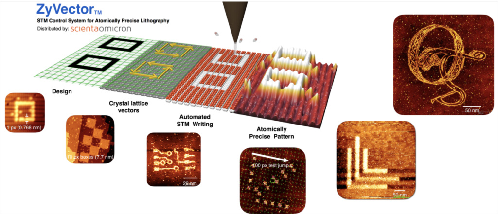 |
The Zyvex Creep and Hysteresis Correction Controller. Live tip position control for fast settling times after landing, and precise motion across the surface. Click here for more information. | 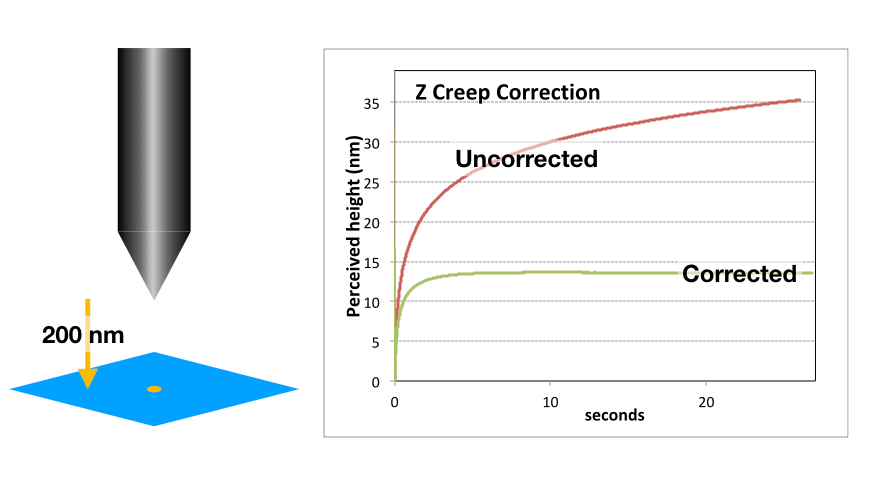 |
The 61st International Conference on Electron, Ion and Photon Beam Technology and Nanofabrication
23rd EIPBN Bizarre/Beautiful Micrograph Contest
“A good Micrograph is worth more than the MegaByte it consumes.”
Entries Presented by Dr. John Randall – Zyvex Labs
The rules include the following:
• Entries have to be of a single image taken with a microscope and should not be significantly altered.
• There is no restriction with respect to the subject matter.
• Electron and ion micrographs have to be black and white.
In 2018, 77 entries were submitted. The entries came from: Australia, Austria, Canada, Finland, France, Italy, Germany, Switzerland, United Kingdom (Scotland), and the United States.
The judges were:
-
Judge Judy – Long suffering spouse of Don Tennant
-
Stella Pang – City University of Hong Kong
-
Hank Smith – MIT
The Judges exercised their prerogative to liberally interpret the award categories, and change the micrograph titles if it pleased them.
There were 7 awards from the judges:
- Grand Prize
- Best Electron Micrograph
- Best Photon Micrograph
- Best Ion Micrograph
- Best STM Micrograph
- Most Bizarre Micrograph
- 3-Beamers Choice Award
There were 6 honorable mentions also given.
All 2018 Entries (with original titles)
GRAND PRIZE
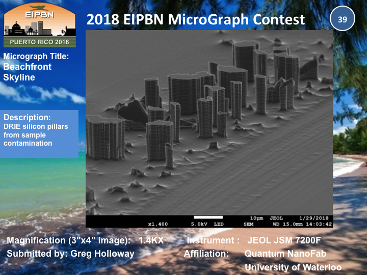
Title: Beachfront Skyline
Description: DRIE silicon pillars from sample contamination
Magnification: 1.4KX
Instrument: JEOL JSM 7200F
Submitted by: Greg Holloway
Affiliation: Quantum NanoFab, University of Waterloo
BEST ELECTRON MICROGRAPH
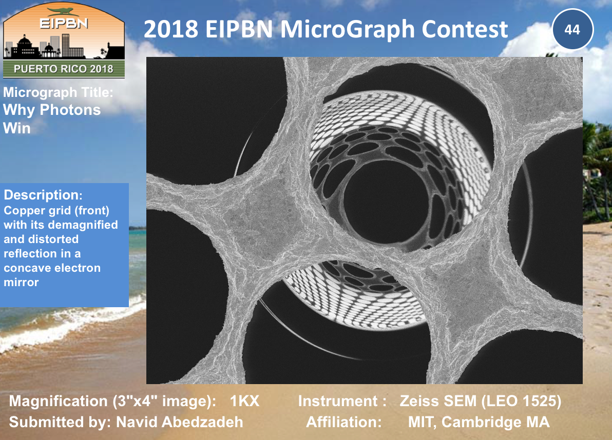
Title: Why Photon Wins
Description: Copper grid (front) with its demagnifiedand distorted reflection in a concave electron mirror
Magnification: (3″x4″ image): 1KX
Instrument: Zeiss SEM (LEO 1525)
Submitted by: Navid Abedzadeh
Affiliation: MIT, Cambridge MA
BEST PHOTON MICROGRAPH
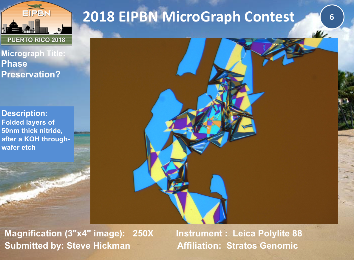
Title: Phase Preservation?
Description: Folded layers of 50nm thick nitride, after a KOH through-wafer etch
Magnification: (3″x4″ image): 250X
Instrument: Leica Polylite88
Submitted by: Steve Hickman
Affiliation: StratosGenomic
Best Ion Micrograph
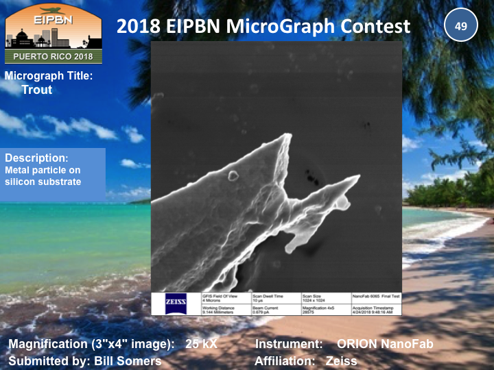
Title: Trout
Description: Metal particle on silicon substrate
Magnification: (3″x4″ image): 25KX
Instrument: ORION NanoFab
Submitted by: Bill Somers
Affiliation: Zeiss
Best STM Micrograph
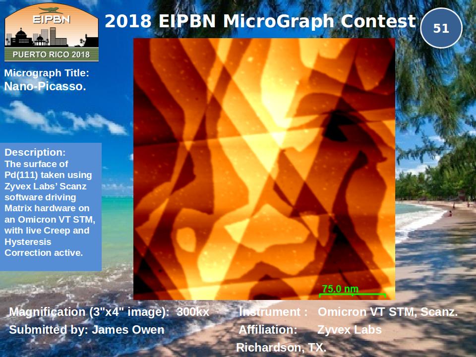
Title: Nano-Picasso
Description: The surface of Pd(111) taken using ZyvexLabs’ Scanzsoftware driving Matrix hardware on an Omicron VT STM, with live Creep and Hysteresis Correction active.
Magnification: (3″x4″ image): 300KX
Instrument: Omicron VT STM, Scanz
Submitted by: James Owen
Affiliation: Zyvex Labs, Richardson, TX
Most Bizarre Micrograph
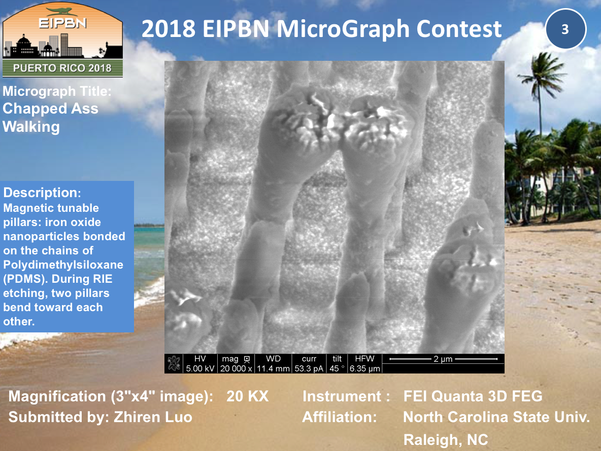
Title: Chapped Ass Walking
Description: Magnetic tunable pillars: iron oxide nanoparticles bonded on the chains of Polydimethylsiloxane (PDMS). During RIE etching, two pillars bend toward each other.
Magnification: (3″x4″ image): 20KX
Instrument: FEI Quanta 3D FEG
Submitted by: Zhiren Luo
Affiliation: North Carolina State University, Raleigh, NC
3-Beamers Choice
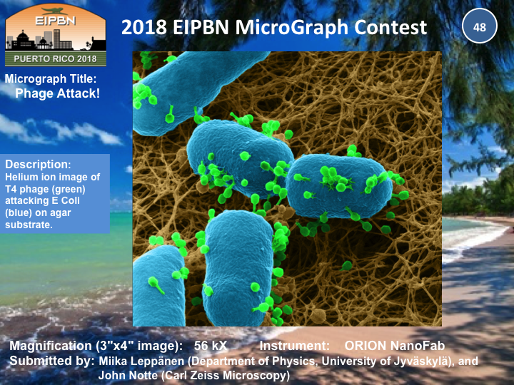
Title: Phage Attack!
Description: Helium ion image of T4 phage (green) attacking E Coli (blue) on agar substrate.
Magnification: (3″x4″ image): 56 KX
Instrument: ORION NanoFab
Submitted by: Miika Leppänen & John Notte
Affiliation: Department of Physics, University of Jyväskylä & Carl Zeiss Microscopy
Honorable Mention
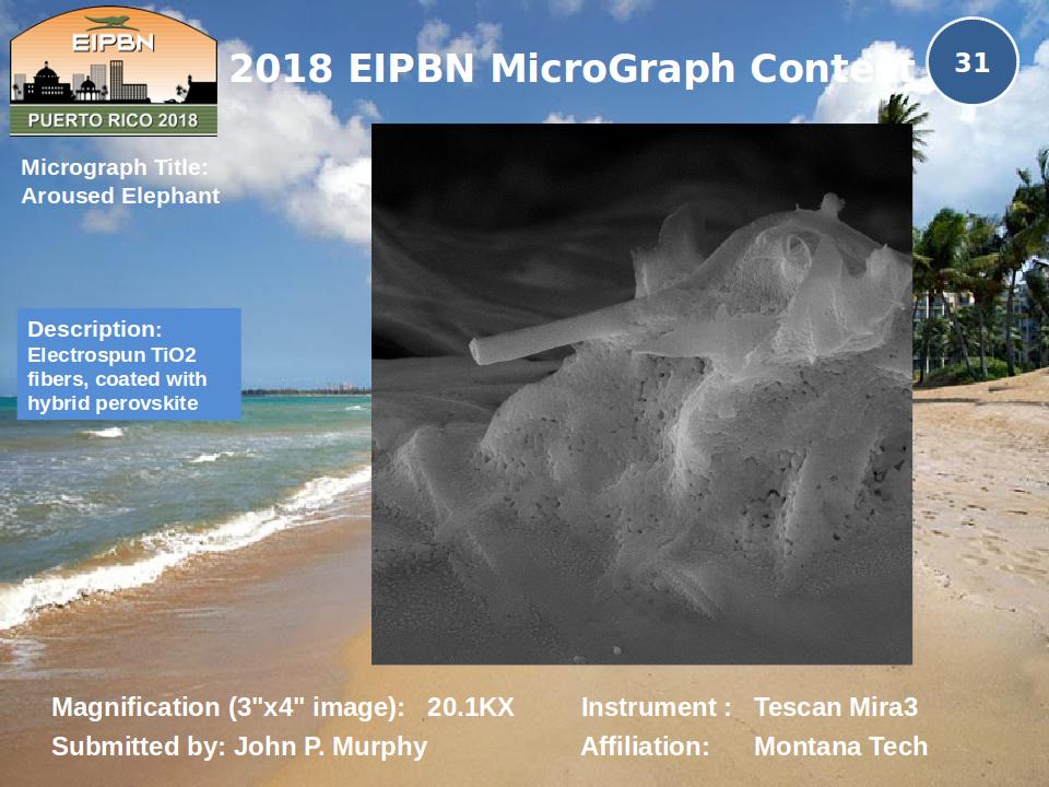
Title: Aroused Elephant
Description: Electrospun TiO2 fibers, coated with hybrid perovskite
Magnification: (3″x4″ image): 20.1 KX
Instrument: TescanMira3
Submitted by: John P. Murphy
Affiliation: Montana Tech
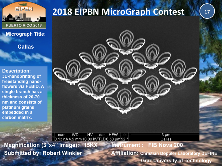
Title: Callas
Description: 3D-nanoprinting of freestanding nano-flowers via FEBID. A single branch has a thickness of 20-70 nm and consists of platinum grains embedded in a carbon matrix.
Magnification: (3″x4″ image): 15KX
Instrument: FIB Nova 200
Submitted by: Robert Winkler
Affiliation: Christian Doppler Laboratory DEFINE; Graz University of Technology
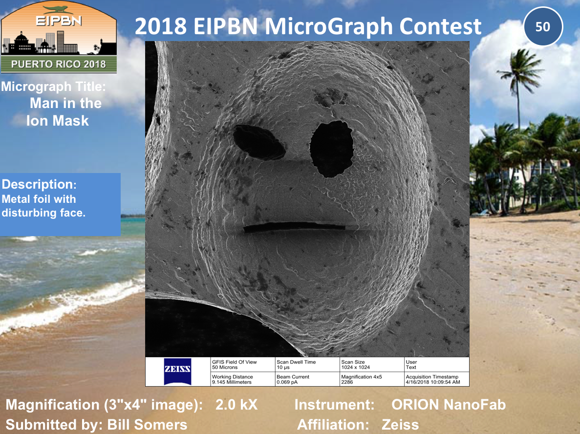
Title: Man in the Ion Mask
Description: Metal foil with disturbing face.
Magnification: (3″x4″ image): 2.0KX
Instrument: ORION NanoFab
Submitted by: Bill Somers
Affiliation: Zeiss
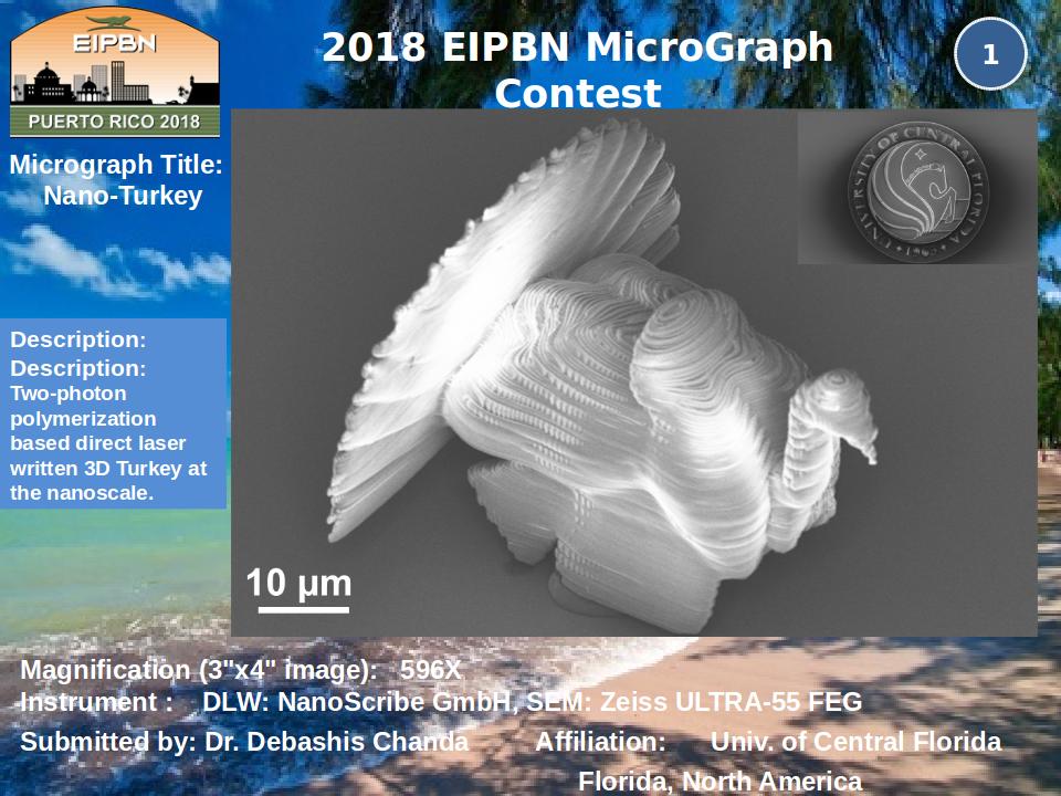
Title: Nano-Turkey
Description: Two-photon polymerization based direct laser written 3D Turkey at the nanoscale.
Magnification: (3″x4″ image): 596X
Instrument: DLW: NanoScribeGmbH, SEM: Zeiss ULTRA-55 FEG
Submitted by: Dr. Debashis Chanda
Affiliation: University of Central Florida
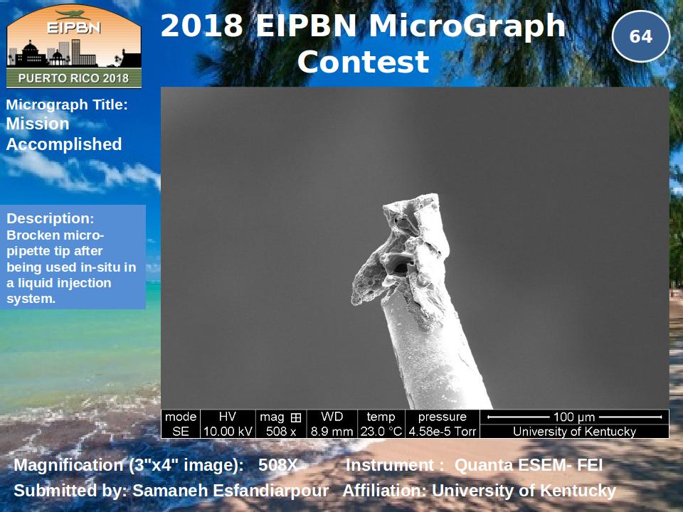
Title: Mission Accomplished
Description: Brocken micro-pipette tip after being used in-situ in a liquid injection system.
Magnification: (3″x4″ image): 508X
Instrument: Quanta ESEM- FEI
Submitted by: SamanehEsfandiarpour
Affiliation: University of Kentucky
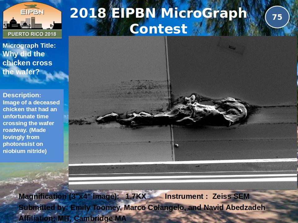
Title: Why did the chicken cross the wafer?
Description: Image of a deceased chicken that had an unfortunate time crossing the wafer roadway. (Made lovingly from photoresist on niobium nitride)
Magnification: (3″x4″ image): 1.7KX
Instrument: Zeiss SEM
Submitted by: Emily Toomey, Marco Colangelo, and NavidAbedzadeh
Affiliation: MIT, Cambridge MA