←2014 |
2016→ |
The 59th International Conference on Electron, Ion and Photon Beam Technology and Nanofabrication
21st EIPBN Bizarre/Beautiful Micrograph Contest
“A good Micrograph is worth more than the MegaByte it consumes.”
Entries Presented by Dr. John Randall – Zyvex Labs
The rules include the following:
• Entries have to be of a single image taken with a microscope and could not be significantly altered.
• There is no restriction with respect to the subject matter.
• Electron and ion micrographs have to be black and white.
In 2015, 118 entries were submitted. Including:
• 88 Electron Micrographs (SEM and TEM)
• 20 Ion Micrographs
• 8 Photon (optical) Micrographs
• 1 Video Micrographs
• 1 Scanning Probe Micrograph
The entries came from: US, China, Canada, Germany, Austria, Hong Kong, UK, Switzerland.
The panel of judges who selected the award winners were:
• Don Tennant – Operations Director of Cornell’s Infamous National Nanofabrication Facility. Originator of Tennant’s Law which has defined the trade-offs in Semiconductor lithography for the last two centuries. One of the oldest 3-Beamers still in captivity.
• Dr. Deirdre Olynick – Staff Scientist, Molecular Foundry Lawrence Berkeley National Laboratory, NanoFabrication guru, and soon to be MBA.
• Judy Tennant -Long suffering spouse of Don Tennant. Veteran of many 3 Beams and Micrograph contests
The Judges exercised their prerogative to liberally interpret the award categories, and change the micrograph titles if it pleased them.
There were six awards:
• Grand Prize
• Most Uplifting
• Most Bizzare
• 3 Beamer’s Choice
• Best Electron Micrograph
• Best Photon Micrograph
There were 8 Honorable Mentions also given.
All 2015 Entries (with original titles)
BEST ELECTRON MICROGRAPH
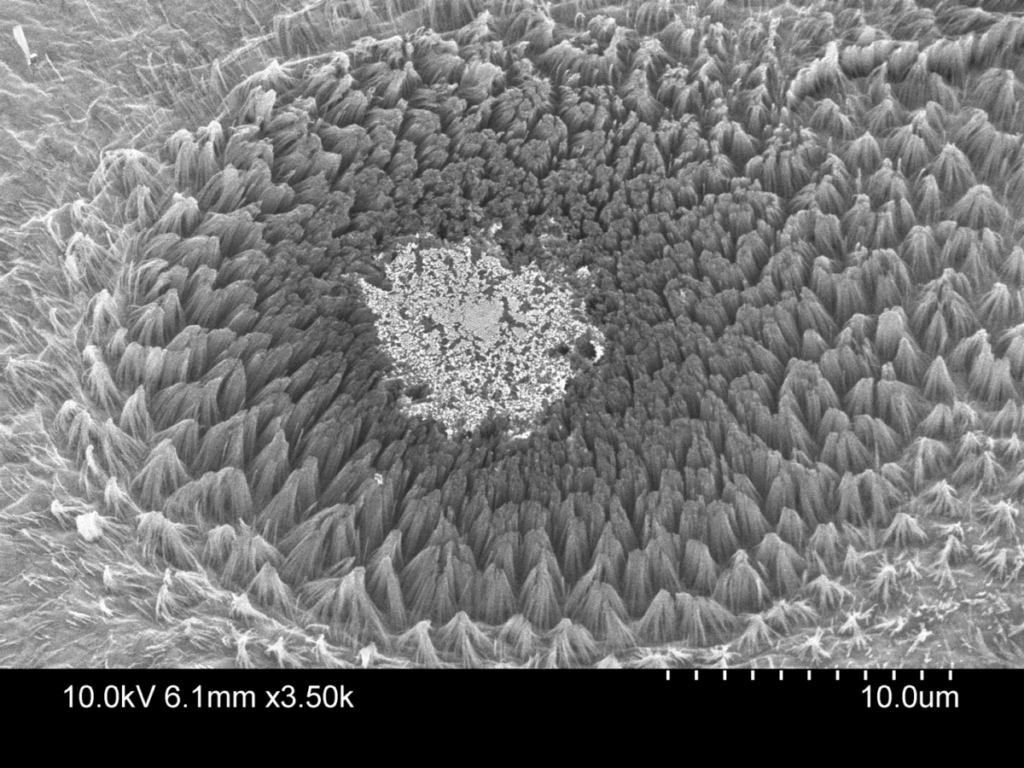
Title: Le Fleur Triumph de Catastrophe
Description: Silicon nanowires etched from silicon wafer by metal assisted chemical etching. Center is gold dots that failed to lift off
Magnification: (3″x 4″ image): 3.5KX
Instrument: Hitachi S-4800 SEM
Submitted by: Kyle Jacobs
Affiliation: University of Illinois (Illinois, North America)
BEST PHOTON PRIZE
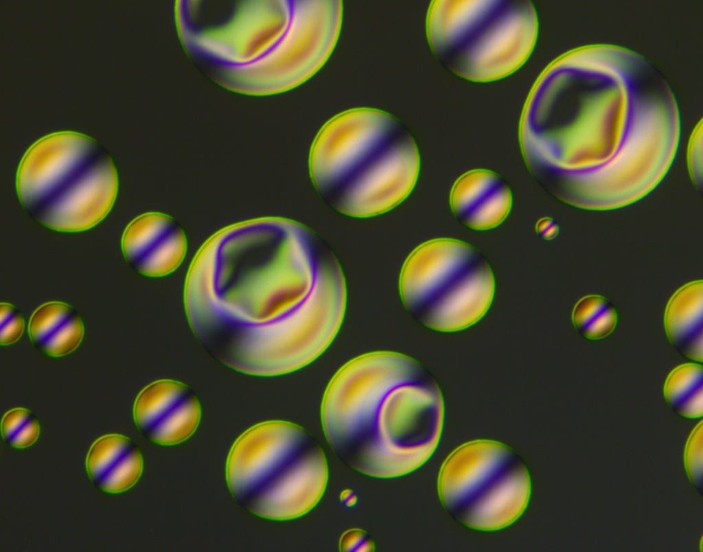
Title:Angry Little Spheriblobs
Description: Multilayers of SiO2 and SiN caused the underlying gold film to bubble up in a beautiful way.
Magnification: (3″x 4″ image): ~120X
Instrument: Nikon Optiphot (DIC imaging)
Submitted by: Rick Bojko, Mark Brunson
Affiliation: Washington Nanofab Facility,U of Washington (Seattle,WA)
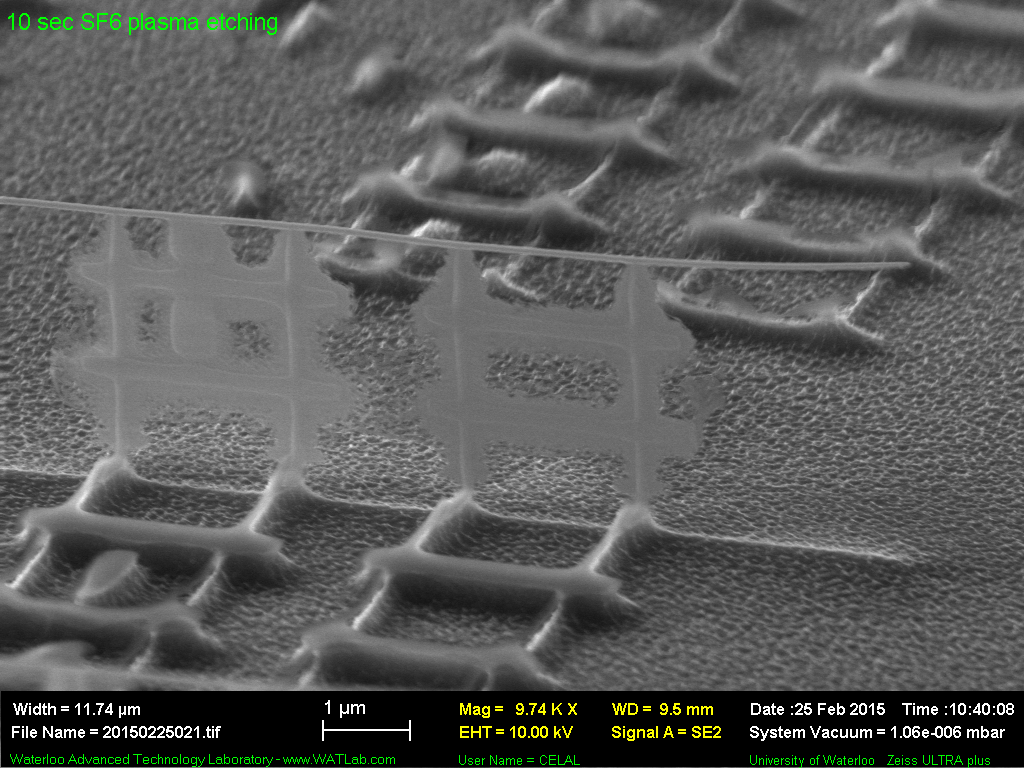
3 BEAMER’S CHOICE AWARD

Title: What you do when spring comes to land: Tulip garden
Description: Silicon nano pillars with metallic shells after RIE process
Magnification: (3″ x 4″ image): 20KX
Instrument: Zeiss Ultra plus FESEM
Submitted by: Celal Con
Affiliation:University of Waterloo (Waterloo, Canada)
BEST aSPIREing MIRCROGRAPH
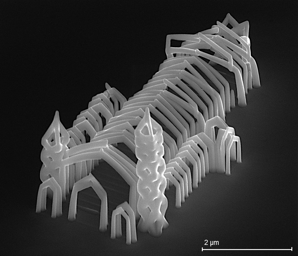
Title: “Herz-Jesu-Cathedral” Graz/Austria
Description: Miniature model of gothic cathedral “Herz-Jesu” in Graz/Austria
Magnification: (3″x 4″ image): 15KX
Instrument: FEI Fib Nova200
Submitted by: Robert Winkler
Affiliation: Graz Centre for Electron Microscopy (Austria)
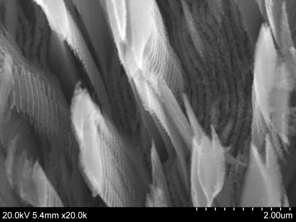
MOST BIZARRE MICROGRAPH
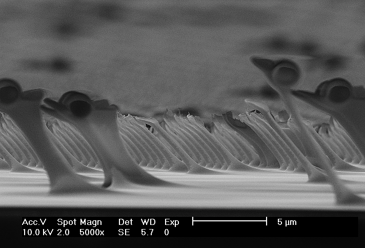
Title: Flying dragons crossing clouds
Description: Pulled up polystyrene during stamp removal
Magnification: (3″x 4″ image): 5KX
Instrument: FEI/Philips XL 30S FEG
Submitted by: Alexander Nöll
Affiliation: University of Wuppertal (Germany)
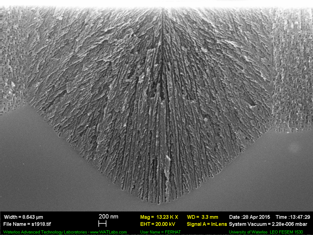
MOST UPLIFTING MICROGRAPH
Title: Ship Happens
Description: Line array and grating on sidewall of Silicon nanowire.
Magnification (3″x 4″ image): 10KX
Instrument: Zeiss Ultra plus FESEM
Submitted by: Celal Con
Affiliation: University of Waterloo (Waterloo,Canada)
GRAND PRIZE
Title: Judy’s Favorite
Description: Silver nano-ribbon assisted chemically etched silicon. Ribbed pillars are silicon, silver ribbons seen at bottom
Magnification: (3″x 4″ image): 20KX
Instrument: Hitachi S-4800 SEM
Submitted by: Kyle Jacobs
Affiliation: University of Illinois (Illinois, North America)
HONORABLE MENTION
Title: The Big Little Big Island
Description: 10 nm pores formed by electrochemical etching under different directions.
Magnification: (3″x 4″ image): 13.23KX
Instrument: LEO FESEM 1530
Submitted by: Ferhat Aydinoglu
Affiliation: University of Waterloo (Waterloo, Canada)
HONORABLE MENTION
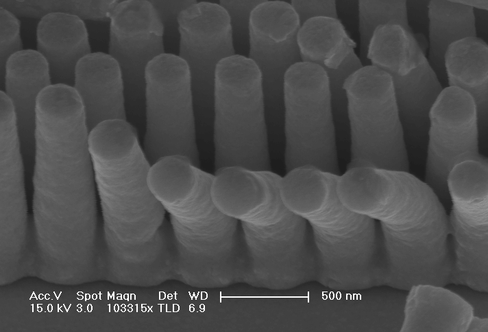
Title: Than’Q very much!
Description: Sub-wavlength gold pillars made a deep bow during electroplating process.
Magnification: (3″x 4″ image): 103KX
Instrument: ZEISS SIGAMA HD
Submitted by: Yaqi Ma
Affiliation: Fudan University (China)
HONORABLE MENTION
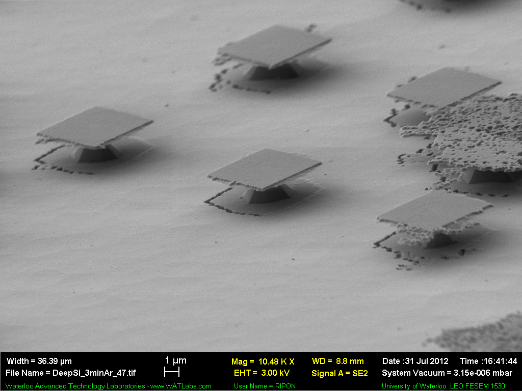
Title: Graduates buried in the sand.
Description: Umbrellas, playing with electron shine!
Magnification: (3″x 4″ image): 11KX
Instrument: Leo FESEM 1530
Submitted by: Ripon Dey
Affiliation: University of Waterloo (Waterloo, Canada)
HONORABLE MENTION
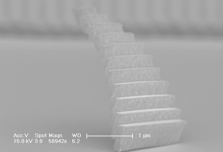
Title: Stairway to Heaven
Description: When resist developing, grating stands up like a ladder.
Magnification: (3″x 4″ image): 59KX
Instrument: FEI-Sirion 200 SEM
Submitted by: Jinhai Shao, Bingrui Lu
Affiliation: Fudan University (China)
HONORABLE MENTION
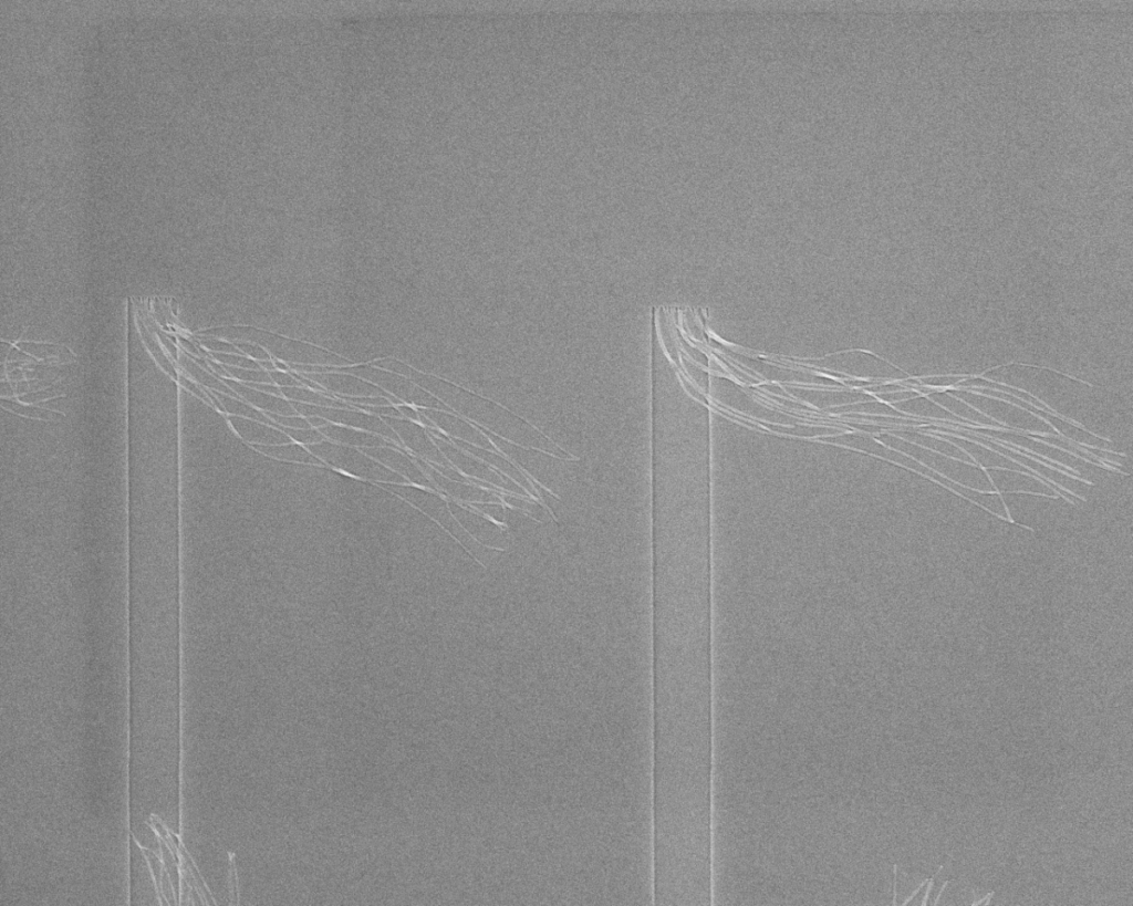
Title: Wind in a Vacuum
Description: Poetic walk in the wind of Nano-land
Magnification: (3″x 4″ image): 10 KX
Instrument: SEM – JEOL JSM7401F
Submitted by: Florian Delechat
Affiliation: University of Montreal (Montreal, QC, Canada)
HONORABLE MENTION
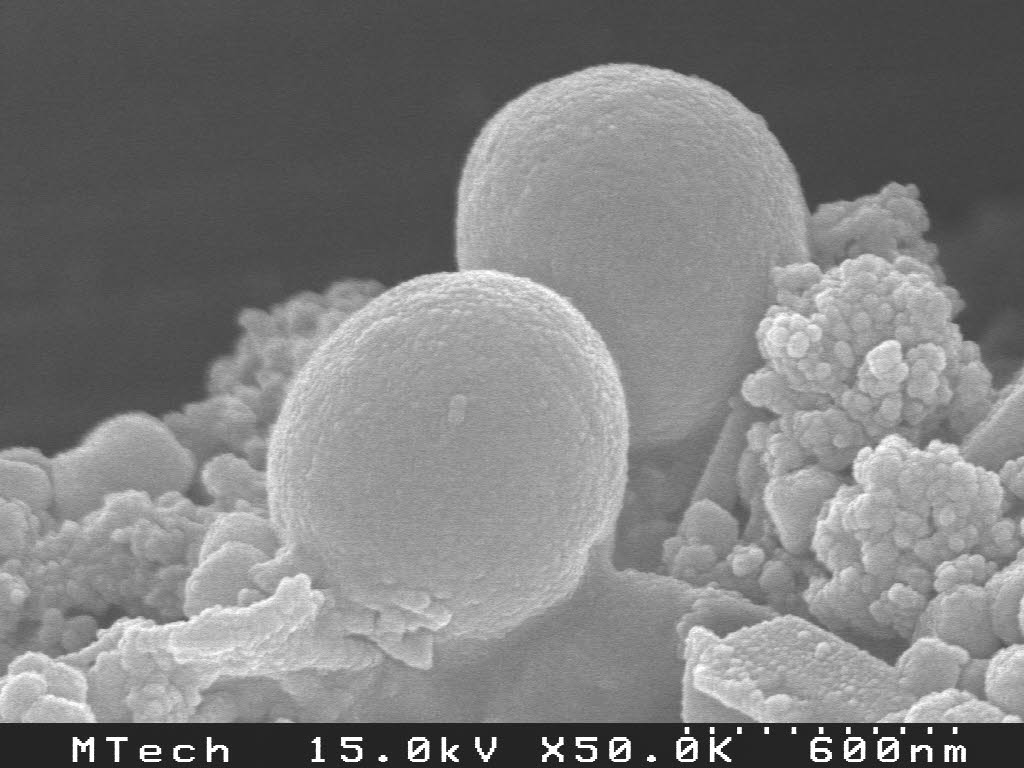
Title: Schwetty Balls
Description: Staphylococcus aureus cells atop gold-doped nanoparticles.
Magnification: (3″x 4″ image): 50KX
Instrument: Hitachi S-4500
Submitted by: Jessica M. Andriolo
Affiliation: Montana Tech:U of M (Butte, MT, North America)
HONORABLE MENTION
Title: Sitteth not on a frustum of a cone
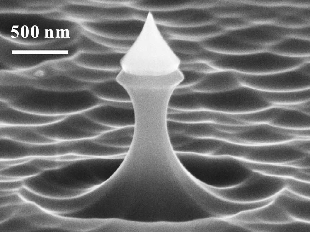
Description: SiO2 coated Si tip supported by the frustum of a cone (85° tilt-view). The lighthouse or the candle floating on the water shows the way to our destination.
Magnification: (3″x 4″ image): 35KX
Instrument: Zeiss SUPRA 60
Submitted by: Zhijun Huang
Affiliation: School of Physics and Engineering, Sun Yat-sen University (China)
HONORABLE MENTION
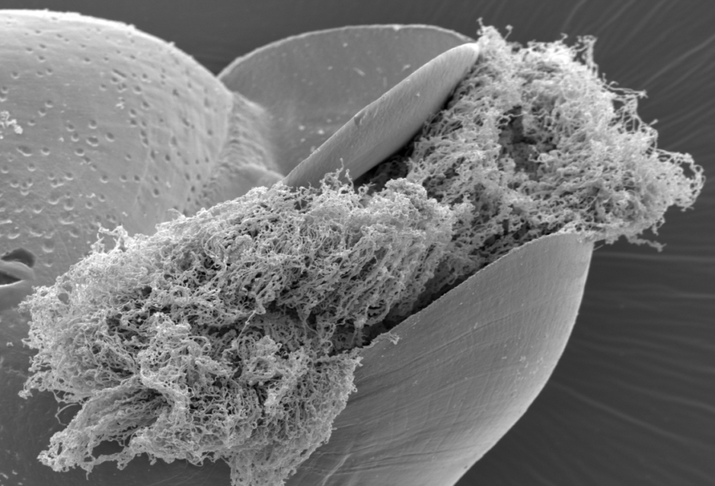
Title: Shredded Snail Taco
Description: Entire nanofiberous cilia of the immature nerite snail are retained, shortened, and wrinkled by using an optional heat fixation step during SEM sample preparation.
Magnification: (3″x 4″ image): 3KX
Instrument: FEI, Philips XL30 ESEM-FEG
Submitted by: Larry Millet
Affiliation: Oak Ridge National Lab (Oak Ridge, TN, North America)