![]()
MNE 2015 Micro Nano Graph Contest
Entries Presented by Dr. John Randall – Zyvex Labs
Sponsored by

|
|
Entries Presented by Dr. John Randall – Zyvex Labs |
Sponsored by |
In 2015, 104 entries received from 21 countries and 5 continents.
RULES
• Entries have to be of a single image taken with a microscope and not significantly altered.
JUDGES
•Nedyalka Panova –Art and Science Visual Artist - Univ. of Cork, Ireland AWARDS:
• First Prize
The judges also selected 12 Honorable Mentions. COMMENTS:
Winners and Honorable Mentions will be displayed in perpetuity at www.ZyvexLabs.com
All 2015 Entries
"Abstract expressionism in polymer microphases"
Description: Phase separation induced by the degradation of the polymer
“nano alphabet soup”
“Hairy pyramids”
Description: InAs nanowires grown on KOH-textured Si (100) substrates. The nanowires grow perpendicular on the side facets of the Si pyramids.
“Atomic Running Track”
Description:
Running track with just the right size for atoms, which is made up of
carbon onion with 0.369 nm gap between each track (layer).
"Monster's Eye"
Description: Dark field image of failed protein crystallization trial on 250 nm thick silicon nitride membrane performed in nanoliter capacity chamber.Deposited protein material clustered around the edges of the membrane and dried out gradually from outside to the center forming reptile like eye effect.
"“The Person and the finger”"
Description:
Magnification (3"x 4" image): 118.06 kX
“Microsphere in the net border!”
Description: Micro metal grid mesh was fabricated by the self-rolling of metal/SiO2 bifilm stress. Polystyrene microparticles were inserted into the micro grid net during self-rolling of the microtube or micro grid net.
"Winter is coming"
Description: Biological cells were cultivated in salt water and this solution was dripped onto a TEM grid for analysis of the cells. The dried NaCl crystals built this snow flake like structures.
"Picasso in the Chamber"
Description: This image is an artefact from the inside of the SEM microscope chamber. It can be tentatively attributed to an electron mirror effect as a result of charge buildup on our electrically isolated sample.
"Awaiting the nano-Titanic"
Description: An islet on Ge surface modified by low energy heavy ions beam.
"Tears of God"
Description: Bubbles caused by chemical etching on Si look like tears.
"TITLE"
Description: Gratings on Si after chemical etching looks like a frightened man who is forbidden to speak.
"TITLE" Nano-Xide Xie
Description: The nano-Xide Xie is fabricated by 3D gray scale e-beam lithography with a wide of 13.26 µm and a height of 18 µm.
Back to Home MNE MicroGraph Contest Home page of the MNE Conference
There were many outstanding micrographs. The work represented in the submitted
micrographs covered a wide range of fields including micro mechanical, photonic,
and integrated circuit fabrication, chemical and dry etching,biological samples,
material science experiments and, of course, e-beam, ion beam, and nano imprint lithography experiments.
• There is no restriction with respect to the subject matter.
• Electron and ion micrographs have to be black and white.
•Vitaliy Guzenko – E-Beam Lithography Master - PSI, Switzerland
•Joshua Ballard – Research Scientist - Zyvex Labs, USA
• Second Prize
• Third Prize
• People's Choice Award
First Prize 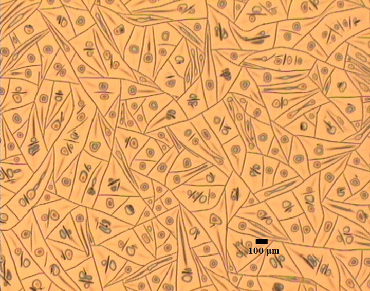
Magnification (3"x 4" image): 10 X
Instrument: OLYMPUS MX51-F
Submitted by: Theodoros Manouras
Affiliation: Institute of Electronic Structure and Laser (IESL), Greece
Second Prize 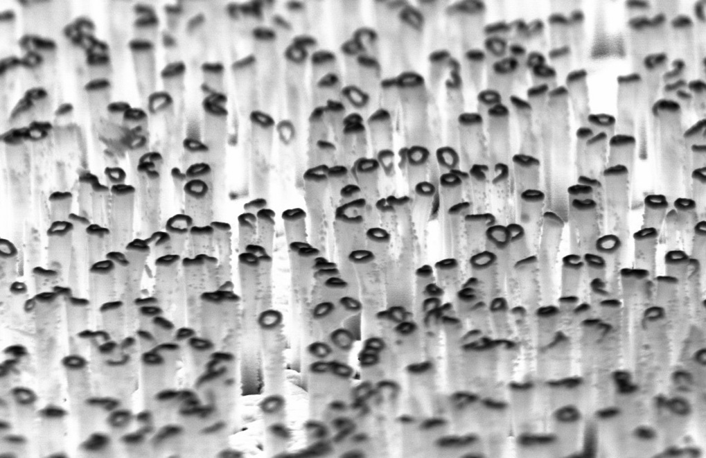
Maskless reactive ion etching of fused silica wi th e-beam deposition 40 nm of Au
Magnification (3"x 4" image): 135.000x
Instrument: SEM ZZeiss Supra
Submitted by: Anil Thilsted & Kristian Sřrensen
Affiliation: DTU Nanotech
Third Prize 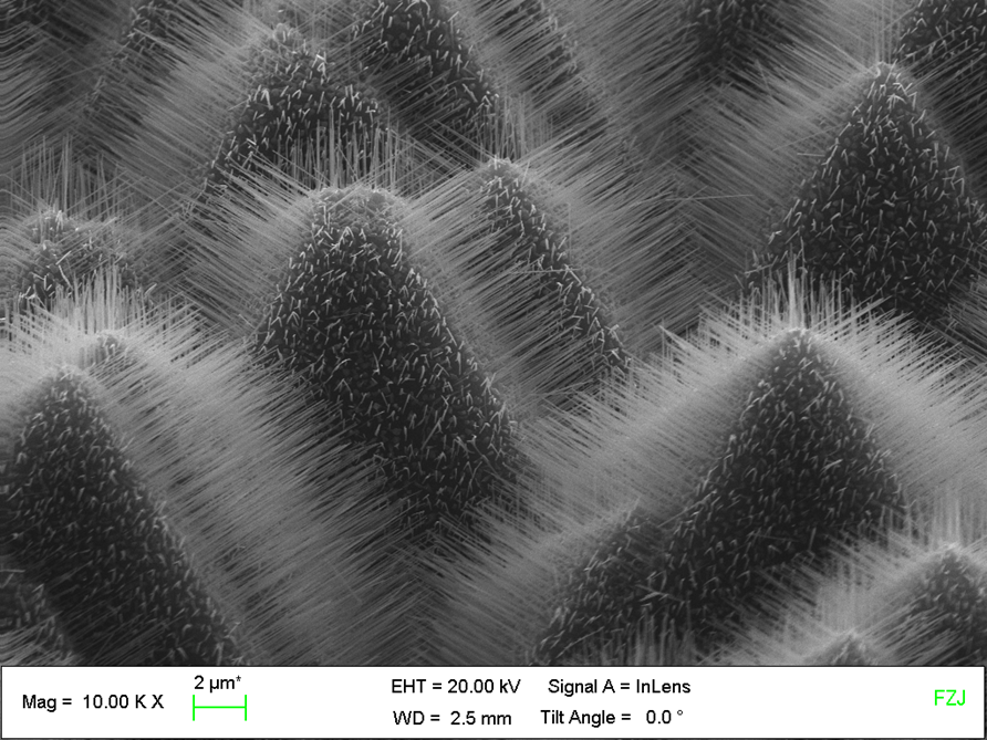
Magnification (3"x 4" image): 10.0 KX
Instrument: Zeiss Gemini 1550
Submitted by: Torsten Rieger
Affiliation: Forschungszentrum Jülich
MNE People's Choice Award - The Judges also selected this micrograph for an honorable meniton award. 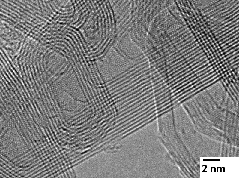
Magnification (3"x 4" image): 500 KX
Instrument: JEOL JEM2100
Submitted by: Tso-Fu Mark Chang
Affiliation: Tokyo Institute of Technology
Honorable Mention 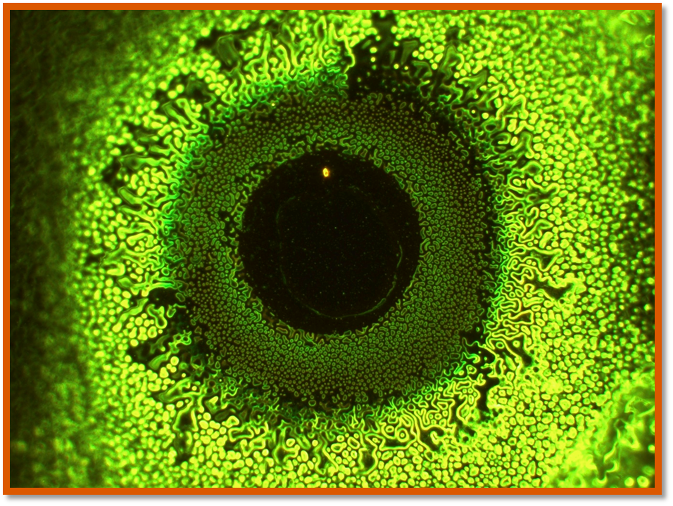
Magnification (3"x 4" image): 800X
Instrument: INM 20 Leica microscope
Submitted by: Nadia Opara
Affiliation: Paul Scherrer Institute, Switzerland
Honorable Mention 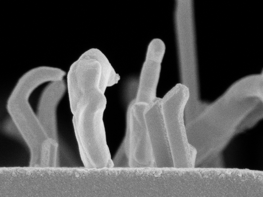
Instrument: Zeiss Supra 55 VP
Submitted by: Robert Kirchner
Affiliation: Paul Scherrer Institute, Switzerland
Honorable Mention 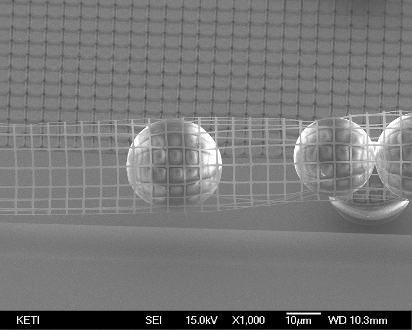
Magnification (3"x 4" image): 1.0 KX
Instrument: JEOL FESEM (JEOL JSM-7000F)
Submitted by: Kook-Nyung Lee
Affiliation: Korea Electronics Technology Institute
Honorable Mention
Magnification (3"x 4" image): 508 X
Instrument: FEI HeIios NanoLab 650 Dualbeam
Submitted by:Thomas Loeber
Affiliation: NSC, TU Kaiserslautern (Kaiserslautern,Germany)
Honorable Mention 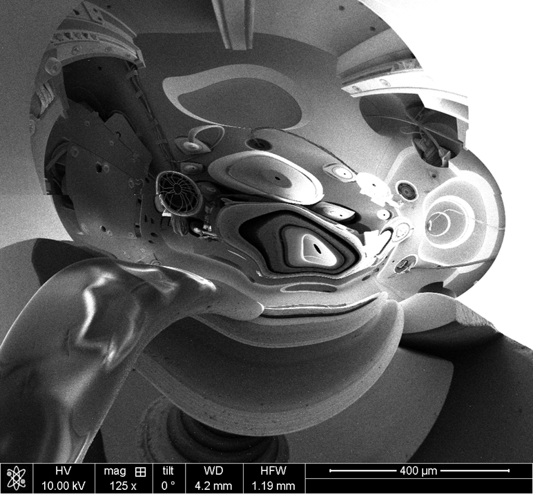
Magnification (3"x 4" image): 125X
Instrument: FEI Helios Nanolab 600
Submitted by:Angelo Accardo
Affiliation: Istituto Italiano di Tecnologia
Honorable Mention 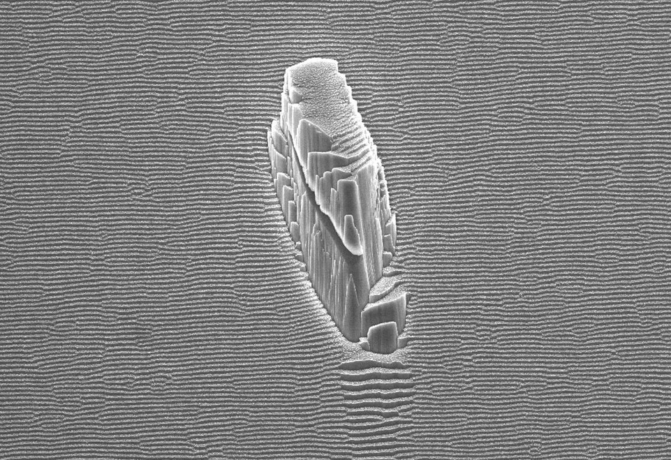
Magnification (3"x 4" image): 3.0 KX
Instrument: SEM Jeol JSM7401F
Submitted by:Erica Iacob
Affiliation: Fondazione Bruno Kessler
Honorable Mention 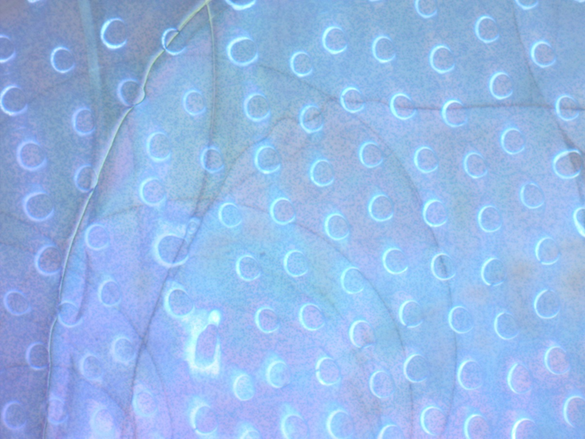
Magnification (3"x 4" image): 1.0 KX
Instrument: Zeiss optical microscope
Submitted by: Xin Li
Affiliation: Fudan University, China
Honorable Mention 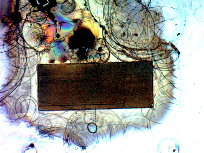
Magnification (3"x 4" image): 1.0 KX
Instrument: Zeiss optical microscope
Submitted by: Xin Li
Affiliation: Fudan University, China
Honorable Mention 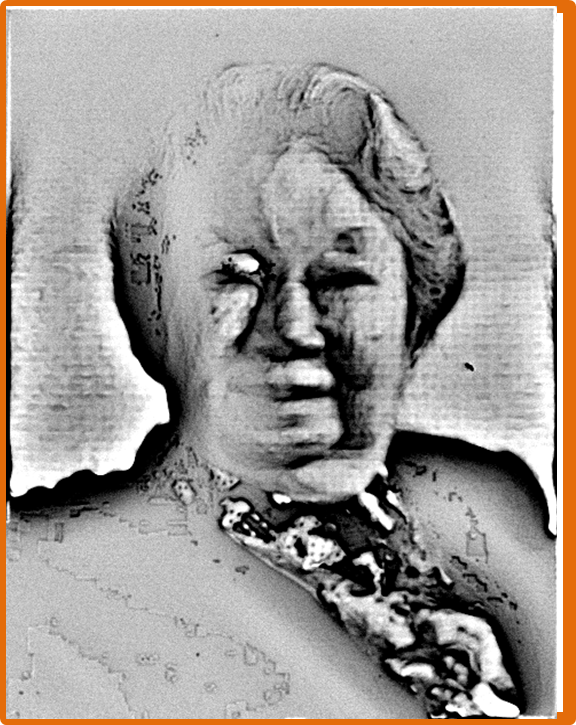
Magnification (3"x 4" image): 4.0 KX
Instrument: JEOL 6300
Submitted by: Chen Xu
Affiliation: Fudan University